Download

1 / 24
260 likes | 565 Vues
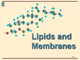
01.20.10 Lecture 4 Cellular Building Blocks: Lipids and Membranes. Membranes form many compartments in the cell. Biological membranes are composed of a lipid bilayer. Membrane lipids are amphipathic molecules. Membrane lipids are amphipathic

E N D
01.20.10Lecture 4 Cellular Building Blocks: Lipids and Membranes
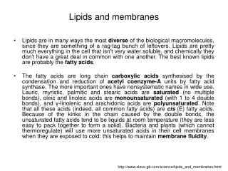
Membrane lipids are amphipathic molecules • Membrane lipids are amphipathic • Hydrophilic heads (polar) form hydrogen bonds with water • Hydrophobic tails (non-polar) are excluded by water molecules
Membrane lipids Phosphatidylcholine is the most abundant phospholipid in cell membranes
Packing arrangements of lipid molecules in an aqueous environment Cone-shaped lipid molecules for micelles, cylinder-shaped lipids form bilayers
Phospholipid bilayers spontaneously close to form a sealed compartment
The membrane bilayer is a fluid • Lateral diffusion occurs rapidly within the plane of the membrane • Individual phospholipids may rotate axially • Flip-flopping from one side to the other is very rare as it is energetically unfavorable
The membrane bilayer is a fluid: FRAP • A fluorescent probe is used to label membrane proteins • The probe is destroyed in a small region using intense laser light • Fluorescence microscopy is used to observe behavior of the unbleached probe
The membrane bilayer is a fluid: single molecule imaging • Fluorescence microscopy of single GFP-labeled membrane proteins • Diffusive movement within the plane of the lipid bilayer
The membrane bilayer is a fluid: laser tweezers Beads trapped in a laser may be used to exert pulling forces on membranes
The composition of a membrane regulates the degree of its fluidity • Membrane lipids with fatty acyl side chains that are saturated (no double bonds) pack tightly in the membrane and make it less fluid • Lipids that are unsaturated (1, 2, or 3 double bonds) pack loosely and make it more fluid
The composition of a membrane regulates the degree of its fluidity The presence of cholesterol in the membrane stiffens the bilayer making it more rigid
Cellular membranes are asymmetric • All lipids are synthesized on the cytosolic surface of the ER • Lipids in the outer leaflets are transported there by flippases • Continuity between organelle lumen & extracellular space
Membrane proteins • Proteins compose ~50% of the membrane • ~1 protein:50 lipid molecules • Membrane proteins perform many functions
Membrane proteins associate with the bilayer in different ways • Transmembrane proteins span the bilayer • Peripheral membrane proteins associate with one side
Transmembrane proteins usually span the bilayer using alpha-helices
Some membrane proteins use beta-sheets to cross the bilayer • Beta-sheets arranged in this cylindrical conformation are known as a “beta-barell” • Hydrophilic amino acid residues face towards the pore, hydrophobics face the bilayer
The cytoplasmic side of the membrane is called the cell cortex • Meshwork of transmembrane proteins and filaments (spectrin) • Mechanical support for the membrane and cell shape
The extracellular surface of the membrane is coated with carbohydrate
Extracellular glycoproteins perform numerous functions • Carbohydrate layer protects cells from chemical and mechanical damage • Different cell types present different combinations of glycoproteins and proteoglycan on their surface - molecular signature • Information in the carbohydrate layer aids in cell-cell recognition and communication
Cells use different mechanisms to restrict membrane protein movements • By tethering to elements inside of the cell (cortex) • By tethering to elements outside of the cell • By interacting with proteins on the surface of another cell • By diffusion barriers established within polarized cells