Download
1 / 30
300 likes | 419 Vues
This resource explores the foundational aspects of biology, focusing on the cellular composition of all living organisms. It highlights the differences between unicellular organisms, such as bacteria and yeast, and multicellular organisms, including plants and animals. Students will learn about the historical significance of cell theory, key scientists like Robert Hooke and Louis Pasteur, and the importance of microscopy in understanding cellular life. By comparing and contrasting these two types of organisms, students gain insights into the complexity and coordination of life.

E N D
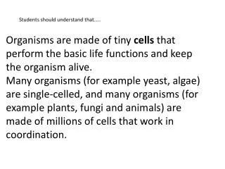
Students should understand that….. Organisms are made of tiny cells that perform the basic life functions and keep the organism alive. Many organisms (for example yeast, algae) are single-celled, and many organisms (for example plants, fungi and animals) are made of millions of cells that work in coordination.
Students should be able to…. Compare and contrast living organisms that are single celled with multicellular organisms.
All living things are made of cells • The cell is the smallest unit that can perform the basic activities of life.
Unicellular Organisms • Unicellular organisms are those that contain just one single cell. • Single-celled organisms have been on Earth for 3.8 billion years. • Although they are only one cell, they are complex.
Unicellular Organism bacteria
protozoa Unicellular Organisms
Unicellular Organisms unicellular algae
Unicellular Organisms unicellular fungi= yeasts
Plants Multicellular Organisms Multicellular organisms are those that are made up of more than one cell. They have specialized cells that, together, meet the basic needs Animals Multicellular Algae Multicellular Fungi
The microscope led to the discovery of the cell (1660s-1670s) Robert Hooke
1660s-1670s Anton Van Leeuwenhoek Anton Van Leeuwenhoek was the first to see and describe bacteria (1674), yeast plants, the teeming life in a drop of water, and the circulation of blood corpuscles in capillaries.
First look at bacteria On September 17, 1683, Leeuwenhoek wrote to the Royal Society about his observations on the plaque between his own teeth, "a little white matter, which is as thick as if 'twere batter." He repeated these observations on two ladies (probably his own wife and daughter), and on two old men who had never cleaned their teeth in their lives. Looking at these samples with his microscope, Leeuwenhoek reported how in his own mouth: "I then most always saw, with great wonder, that in the said matter there were many very little living animalcules, very prettily a-moving. The biggest sort. . . had a very strong and swift motion, and shot through the water (or spittle) like a pike does through the water. The second sort. . . oft-times spun round like a top. . . and these were far more in number." In the mouth of one of the old men, Leeuwenhoek found "an unbelievably great company of living animalcules, a-swimming more nimbly than any I had ever seen up to this time. The biggest sort. . . bent their body into curves in going forwards. . . Moreover, the other animalcules were in such enormous numbers, that all the water. . . seemed to be alive." These were among the first observations on living bacteria ever recorded.
Schleiden, Schwann and Virchow 1850s Cell Theory "all living things are composed of living cells" Schwann: Discovered that all animals are made up of cells. "All living cells arise from pre-existing cells". Virchow: Discovered that cells only come from other cells. Schleiden: Announced that all plants are composed of cells. Virchow
Louis Pastuer He was able to demonstrate that organisms such as bacteria were responsible for souring wine and beer (he later extended his studies to prove that milk was the same). The bacteria could be removed by heating wine, beer, milk, or vinegar briefly, thereby sterilizing—or 'pasteurizing'—the substances.
Louis Pasteur helped prove there was no spontaneous creation of cells from non-living matter • scientists believed living things could spontaneously generate. people believed that mice can arise in the pot with grain, shut by dirty shirt.
Spontaneous Generation? Or Not? Heat- kills bacteria Left open- bacteria appear conclusion: spontaneous generation is true Stoppered – No bacteria grow = no spontaneous generation Argument – Hey! Nothing grows with no air! How do we resolve this question?
Pasteur's Experiment Trying to prove the living organisms that grew came from outside, as spores on dust, rather than spontaneously generated within the broth. Flask changed into a long S shape= prevented microorganisms in the air from easily entering the flask, yet allowed some air interchange. If the swan neck was broken, microbes readily entered the flask and grew.
Pasteur & Vaccinations The principle of vaccination or immunization is based on the property of 'memory' of the immune system. A vaccine is defined as an inoculation containing germs in dead, weakened or virulent form or modified toxins which when injected inside the body, stimulates the defensive system of the body to produce antibodies. This creates an immunity inside the body of an organism against that particular disease.
Germ Theory • Beverage contamination led Pasteur to the idea that micro-organisms infecting animals and humans cause disease.
Cell Theory 1. All living organisms are composed of one or more cells. 2. The cell is the basic unit of structure, function, and organization in all organisms. 3. All cells come from preexisting cells.
pollen grains from a variety of common plants Scanning electron micrograph of a sipunculanpelagosphera larva Gills of a fish, the mudskipper (Periophthalmusargentilineatus) lung trachea epithelium
Comparing TEM and SEM Transmission Electron Microscopes • The transmission electron microscope (TEM), the first type of EM, has many commonalities with the optical microscope and is a powerful microscope, capable of producing images 1 nanometer in size. • They require high voltages to increase the acceleration speed of electrons, which, once they pass through the sample (transmission), increase the image resolution. • The 2-d, black and white images produced by TEMs can be seen on a screen or printed onto a photographic plate. • Although recent innovations in software help to minimize, TEM resolution is hampered by spherical and chromatic aberrations. • The TEM is a popular choice for nanotechnology as well as semiconductor analysis and production. Scanning Electron Microscopes • Reflecting light microscopes are the optical counterpart to scanning electron microscopes (SEM) and produce similar data. • SEMs are primarily used to obtain topographical information. • In this type of EM, a series of solenoids pulls the beam back and forth across the sample, systematically scanning the surface; it detects secondary electrons emitted from the surface and produces an image. • Although SEMs are approximately 10 times less powerful than TEMs, they produce high-resolution, sharp, black and white 3D images.