Download
1 / 37
370 likes | 643 Vues
Enzyme linked plasma membrane surface receptors Tyr kinases. Growth factors - regulation of proliferation. Endothelial cell growth factor -. ECGF. Epidermal growth factor -. EGF. its receptor coding gene: c erb B. Fibroblast growth factor -. FGH.

E N D
Growth factors - regulation of proliferation Endothelial cell growth factor - ECGF Epidermal growth factor - EGF its receptor coding gene: c erb B Fibroblast growth factor - FGH (endothelial cells, fibroblast, smooth muscle cell, granulosa cell, chondrocyte) Insulin Insulin like growth factor - IGF Interleukins Nerve growth factor - NGF (neurons) Platelet derived growth factor - (PDGF) factor coding gene: c-sis (fibroblast, glia cells, keratinocyte, epithelial cells, endothelial cells, chondrocyte)
Four intracellular pathways cross talk
A Single Signal Can Activate Several Pathways Binding of epidermal growth factor to its receptor activates phospholipase C (PLC) leading to production of diacylglycerol (DAG) and activation of protein kinase C (PKC) Binding of epidermal growth factor to its receptor activates a MAP kinase pathway via ras DAG ras PLC raf PKC ras GTP MAP kinase cascade Multiple effects e.g. Differentiation Proliferation
protein kinases activated through receptors • AGC group: PKA, PKG, PKC, Rac, G-protein kinases • CaMK group: kinases regulated by Ca2+/CaM • CMGC group: cyclin-dependent kinases ERK, MAP, Casein kinase • PTK group: conventional protein tyrosine kinases Src, Abl, Fak, PDGF, IR 5. OPK: Other Protein Kinases appr. 2000 protein kinases in human genome
Protein tyrosine kinases (PTK) • Plasma membrane receptors - receptor tyrosine kinases • dimer formation, autophosphorylation 2. Cytoplasmic tyrosine kinases
Src, is a non-receptor tyrosine kinase Src, is the product of the first proto-oncogene to be characterized. Src kinase -„eldest” Tyr kinase – SH: Src Homology domain
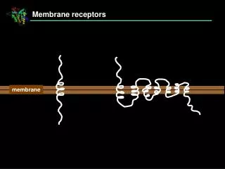
Tyrosine kinases can be cytosolic or integral membrane receptors . Substrate Single Membrane spanning Hydrophobic domain. No membrane-spanning domain
Src homology domains SH2 binding to Tyr P SH3 binding to Pro rich regions Grb2: SH2, SH3 domains – receptor –effector connection Docking proteins: SH2 domain IRS: insulin receptor substrate
Receptor tyrosine kinases The interaction of the external domain of a receptor tyrosine kinase with the ligand, often a growth factor, up-regulates the enzymatic activity of the intracellular catalytic domain, which causes tyrosine phosphorylation of cytoplasmic signaling molecules.
Receptor tyrosine kinases • General Relevance • Amplification by downstream signaling elements greatly amplifies the effects of low levels of tyrosine phosphorylation that are most directly induced by extracellular triggers. Example: PLC and PI3K • Activation of multiple kinases (kinase cascades) including ser/thr as well as tyrosine kinases, is a frequent consequence of these early events. Example: MAP Kinase
Module 1: Figure PDGFR activation Cell Signalling Biology www.cellsignallingbiology.org 2007
Tyr-P docking sites on receptor protein or binding of SH2 proteins to plasma membrane activation of PLC IP3 DAG MAP kinase activation of Ras PI3 - kinase PKB (protein kinase B) Survival inhibition of apoptosis increase in glycogen synthesis stimulation of translation (mTor)
Functionsof receptor tyrosine kinases: • Growth control • Cell-cell recognition • Cell cycle control • Immune responses • Development • Differentiation
Tyrosine Kinases, associated genes and proteins are implicated in developmental defects and cancer • Excessive activation of receptor tyrosine kinases can lead to uncontrolled growth and malignant transformation. • Many defective or viral forms of tyrosine kinases and associated proteins are oncogenic: • v-src • abl • erbB
Module 1: Figure tyrosine kinase-linked receptors Cell Signalling Biology www.cellsignallingbiology.org 2007
Response of the insulin receptor kinase (IRK) to ligand binding • Heterotetramer (2a, 2b) • Insulin binding leads to change in structure (different from other RTKs) • Conformation change activates b-subunit TK activity • b subunit phosphorylates Tyr residues on cytoplasmic domains as well as downstream substrates (IRS)
Three-dimensional structures of the insulin receptor tyrosine kinase (IRK) IRK conformational change upon activation loop phosphorylation. The N-terminal lobe of IRK is colored white and the C-terminal lobe is colored dark grey. The activation loop (green) contains autophosphorylation sites Y1158, Y1162 and Y1163, and the catalytic loop (orange) contains the putative catalytic base, D1132. Also shown are the unbound/bound ATP analog and tyrosine-containing substrate peptide (pink). [Hubbard, EMBO J. 16, 5572 (1997)]
Once Tyr-phosphorylated, the IRK activity triggers a number of signaling pathways • Phosphatidylinositol 3-hydroxy kinase, makes PIP2,PIP3 • Grb2, Sos, activates Ras • Activation of PI-PLC Sos: exchange protein
Module 2: Figure insulin receptor Cell Signalling Biology www.cellsignallingbiology.org 2007
IR (insulin receptor) IRS (insulin receptor substrate) (IRS1, IRS2) IRS dependent phosphatidylinositol 3-kinase (PI3K) PIP3 (PI – 3,4,5 trisphosphate) aPKC (atypical protein kinase C) PKB/Akt Sterol regulatory element binding protein -1c (SREBP-1c) liver FAS (fatty acid synthase) Acetyl CoA carboxylase
On-Off Switches The majority of signals are transient and the response should be proportionately transient too. If you switch the signal on, you need a way of switching it off again. For example, failure to switch off mitogenic signals is one way to induce a tumour. So, what we are looking for are biochemical systems that are capable of rapidly switching between two states. In many signalling systems the ‘on-off’ switch is operated by GTP-binding proteins and/or protein phosphorylation
On-Off Switches – GTP-Binding Proteins GTP-binding proteins come in two flavours, small monomeric GTP-binding proteins (e.g. p21ras) and heterotrimeric G proteins. The basic GTP/GDP binding cycle is the same in both cases. g Exchange of bound GDP for GTP b GTP a INACTIVE GDP GDP a a subunit dissociates from bg GTP ACTIVE Pi Active a subunit can interact with and activate the next step in the signalling pathway a a subunit GTPase activity GTP > GDP+Pi GDP a subunit reassociates with bg
p21ras p21ras p21ras GDP GDP GTP On-Off Switches – GTP-Binding Proteins Ras (p21ras) is a good example of this type of switch. Ras is a small (21 kDa) monomeric protein that binds GTP or GDP and has intrinsic GTPase activity This causes exchange of bound GDP for GTP Guanine nucleotide exchange factor interacts with ras p21ras Activated ras interacts with and activates the next component in the signalling pathway On GTP GDP ACTIVE INACTIVE Pi Ras GTPase stimulated by association with GTPase-activating protein (GAP) Off Intrinsic GTPase activity hydrolyses GTP to GDP and Pi
RTKs can activate the Ras pathway of cellular signaling • Ras is a small G-protein (monomeric 21-kD) • Mutant Ras proteins are unable to dissociate GTP, so they are stuck in the ON or proliferative state: ras (gene) mutations found in 30% of human cancers. • Mutations in Ras-GAPs can lead to disease.
Unlike IRK, most RTKs are present as a monomer in the resting cell membrane
Steps in the activation of Ras by RTKs Raf is a PK that triggers MAP-K pathway Raf SH2 binds RTK, SH3 binds SOS c-fos, c-jun Cell proliferation Ras-GEF
Cascading Kinases Binding of epidermal growth factor to its receptor activates ras Ras activates the serine/threonine kinase raf Erk-1 phosphorylates the transcription factor myc and activates transcription ras raf raf ras GTP GDP ADP ATP Raf phosphorylates and activates the dual-specificity kinase Mek-1 P Mek1 Mek1 Nucleus ATP P Erk1 ATP ADP ADP P P P Erk1 Erk1 Mek-1 phosphorylates the serine/threonine kinase Erk-1 which migrates to the nucleus
Signal transduction of cytokines 1. protein signals (IL - 1 IL - 13) paracrine, autocrine regulation heterodimer cytokine receptors gp130 - common constituent in several receptors signal - receptor complex binding of cytoplasmic tyrosine kinases to the complex (JAK etc.) autophosphorylation (tyr kinases + receptor) SH2 proteins JAK: Janus kinase
Signal transduction of cytokines 2. STAT proteins transcription factors phosphorylation at Tyr dimer formation translocation into nucleus (Ser-P) binding to specific enhancer elements activation of specific genes
Module 2: Figure JAK/STAT function Cell Signalling Biology www.cellsignallingbiology.org 2007
P Nucleus A Simple Signalling System INFg receptor Interferon (IFN) g Activated IFNg receptor recruits JAK kinase Phosphorylation causes STATs to dimerise and migrate to nucleus… …where they initiate transcription ADP ATP JAK phosphorylates STAT monomer STAT transcription factor JAK tyrosine kinase
Module 2: Figure JAK/STAT heterogeneity Cell Signalling Biology www.cellsignallingbiology.org 2007
Module 1: Figure cytokines Cell Signalling Biology www.cellsignallingbiology.org 2007