18 The Endocrine System
1.45k likes | 1.88k Vues
18 The Endocrine System. An Introduction to the Endocrine System. Learning Outcomes 18-1 Explain the importance of intercellular communication, describe the mechanisms involved, and compare the modes of intercellular communication that occur in the endocrine and nervous systems.

18 The Endocrine System
E N D
Presentation Transcript
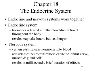
18 The Endocrine System
An Introduction to the Endocrine System • Learning Outcomes • 18-1 Explain the importance of intercellular communication, describe the mechanisms involved, and compare the modes of intercellular communication that occur in the endocrine and nervous systems. • 18-2 Compare the cellular components of the endocrine system with those of other systems, contrast the major structural classes of hormones, and explain the general mechanisms of hormonal action on target organs.
An Introduction to the Endocrine System • Learning Outcomes • 18-3 Describe the location, hormones, and functions of the pituitary gland, and discuss the effects of abnormal pituitary hormone production. • 18-4 Describe the location, hormones, and functions of the thyroid gland, and discuss the effects of abnormal thyroid hormone production. • 18-5 Describe the location, hormone, and functions of the parathyroid glands, and discuss the effects of abnormal parathyroid hormone production.
An Introduction to the Endocrine System • Learning Outcomes • 18-6 Describe the location, structure, hormones, and general functions of the adrenal glands, and discuss the effects of abnormal adrenal hormone production. • 18-7 Describe the location of the pineal gland, and discuss the functions of the hormone it produces. • 18-8 Describe the location, structure, hormones, and functions of the pancreas, and discuss the effects of abnormal pancreatic hormone production.
An Introduction to the Endocrine System • Learning Outcomes • 18-9 Describe the functions of the hormones produced by the kidneys, heart, thymus, testes, ovaries, and adipose tissue. • 18-10 Explain how hormones interact to produce coordinated physiological responses and influence behavior, describe the role of hormones in the general adaptation syndrome, and discuss how aging affects hormone production and give examples of interactions between the endocrine system and other organ systems.
An Introduction to the Endocrine System • The Endocrine System • Regulates long-term processes • Growth • Development • Reproduction • Uses chemical messengers to relay information and instructions between cells
Figure 18-1 Organs and Tissues of the Endocrine System Hypothalamus Pineal Gland Melatonin Production of ADH, oxytocin, and regulatory hormones Parathyroid Glands (located on the posterior surface of the thyroid gland) Pituitary Gland Anterior lobe: Parathyroid hormone (PTH) ACTH, TSH, GH, PRL, FSH, LH, and MSH Posterior lobe: Release of oxytocin and ADH
Figure 18-1 Organs and Tissues of the Endocrine System Organs with Secondary Endocrine Functions See Chapter 21 Heart: Secretes natriuretic peptides. • Atrial natriuretic peptide (ANP) • Brain natriuretic peptide (BNP) Thyroid Gland Thymus: (Undergoes atrophy during adulthood) Secretes thymosins See Chapter 22 Thyroxine (T4) Triiodothyronine (T3) Calcitonin (CT) Adipose Tissue: Secretes • Leptin Adrenal Glands See Chapter 25 Digestive Tract: Secretes numerous hormones involved in the coordination of system functions, glucose metabolism, and appetite Adrenal medulla: Epinephrine (E) Norepinephrine (NE) Adrenal cortex: See Chapters 19 and 26 Kidneys: Secrete • Erythropoietin (EPO) • Calcitriol Cortisol, corticosterone, aldosterone, androgens Gonads: See Chapters 28 and 29 Pancreas (Pancreatic Islets) Testis Testes (male): Androgens (especially testosterone), inhibin Insulin Glucagon Ovaries (female): Estrogens, progestins, inhibin Ovary
18-1 Homeostasis and Intercellular Communication • Direct Communication • Exchange of ions and molecules between adjacent cells across gap junctions • Occurs between two cells of same type • Highly specialized and relatively rare • Paracrine Communication • Uses chemical signals to transfer information from cell to cell within single tissue • Most common form of intercellular communication
18-1 Homeostasis and Intercellular Communication • Endocrine Communication • Endocrine cells release chemicals (hormones) into bloodstream • Alters metabolic activities of many tissues and organs simultaneously
18-1 Homeostasis and Intercellular Communication • Target Cells • Are specific cells that possess receptors needed to bind and “read” hormonal messages • Hormones • Stimulate synthesis of enzymes or structural proteins • Increase or decrease rate of synthesis • Turn existing enzyme or membrane channel “on” or “off”
18-1 Homeostasis and Intercellular Communication • Synaptic Communication • Ideal for crisis management • Occurs across synaptic clefts • Chemical message is “neurotransmitter” • Limited to a very specific area
18-2 Hormones • Classes of Hormones • Hormones can be divided into three groups • Amino acid derivatives • Peptide hormones • Lipid derivatives • Secretion and Distribution of Hormones • Hormones circulate freely or travel bound to special carrier proteins
18-2 Hormones Amino Acid Derivatives Are small molecules structurally related to amino acids Derivatives of Tyrosine: Thyroid hormones Catecholamines Epinephrine, norepinephrine Derivatives of Tryptophan: Dopamine, serotonin, melatonin
18-2 Hormones Peptide Hormones Are chains of amino acids Most are synthesized as prohormones Inactive molecules converted to active hormones before or after they are secreted Glycoproteins Proteins are more than 200 amino acids long and have carbohydrate side chains Thyroid-stimulating hormone (TSH) Luteinizing hormone (LH) Follicle-stimulating hormone (FSH)
18-2 Hormones Peptide Hormones Short Polypeptides/Small Proteins Short chain polypeptides Antidiuretic hormone (ADH) and oxytocin (OXT) (each 9 amino acids long) Small proteins Growth hormone (GH; 191 amino acids) and prolactin (PRL; 198 aminoacids) Includes all hormones secreted by: Hypothalamus, heart, thymus, digestive tract, pancreas, and posterior lobe of the pituitary gland, as well as several hormones produced in other organs
18-2 Hormones Lipid Derivatives Eicosanoids - derived from arachidonic acid, a 20-carbon fatty acid Paracrine factors that coordinate cellular activities and affect enzymatic processes (such as blood clotting) in extracellular fluids Some eicosanoids (such as leukotrienes) have secondary roles as hormones A second group of eicosanoids - prostaglandins - involved primarily in coordinating local cellular activities In some tissues, prostaglandins are converted to thromboxanes and prostacyclins, which also have strong paracrine effects
18-2 Hormones Lipid Derivatives Steroid hormones - derived from cholesterol Released by: The reproductive organs (androgens by the testes in males, estrogens and progestins by the ovaries in females) The cortex of the adrenal glands (corticosteroids) The kidneys (calcitriol) Because circulating steroid hormones are bound to specific transport proteins in the plasma: They remain in circulation longer than secreted peptide hormones
18-2 Hormones • Secretion and Distribution of Hormones • Free Hormones • Remain functional for less than 1 hour • Diffuse out of bloodstream and bind to receptors on target cells • Are broken down and absorbed by cells of liver or kidneys • Are broken down by enzymes in plasma or interstitial fluids
18-2 Hormones • Secretion and Distribution of Hormones • Thyroid and Steroid Hormones • Remain in circulation much longer because most are “bound” • Enter bloodstream • More than 99% become attached to special transport proteins • Bloodstream contains substantial reserve of bound hormones
18-2 Hormones • Mechanisms of Hormone Action • Hormone Receptor • Is a protein molecule to which a particular molecule binds strongly • Responds to several different hormones • Different tissues have different combinations of receptors • Presence or absence of specific receptor determines hormonal sensitivity
18-2 Hormones • Hormones and Plasma Membrane Receptors • Catecholamines and Peptide Hormones • Are not lipid soluble • Unable to penetrate plasma membrane • Bind to receptor proteins at outer surface of plasma membrane (extracellular receptors) • Eicosanoids • Are lipid soluble • Diffuse across plasma membrane to reach receptor proteins on inner surface of plasma membrane (intracellular receptors)
18-2 Hormones • Hormones and Plasma Membrane Receptors • First and Second Messengers • Bind to receptors in plasma membrane • Cannot have direct effect on activities inside target cell • Use intracellular intermediary to exert effects
18-2 Hormones First Messenger Leads to second messenger May act as enzyme activator, inhibitor, or cofactor Results in change in rates of metabolic reactions
18-2 Hormones • Important Second Messengers • Cyclic-AMP (cAMP) • Derivative of ATP • Cyclic-GMP (cGMP) • Derivative of GTP • Calcium ions
18-2 Hormones • The Process of Amplification • Is the binding of a small number of hormone molecules to membrane receptors • Leads to thousands of second messengers in cell • Magnifies effect of hormone on target cell
18-2 Hormones • Down-regulation • Presence of a hormone triggers decrease in number of hormone receptors • When levels of particular hormone are high, cells become less sensitive to it • Up-regulation • Absence of a hormone triggers increase in number of hormone receptors • When levels of particular hormone are low, cells become more sensitive to it
18-2 Hormones • G Protein • Enzyme complex coupled to membrane receptor • Involved in link between first messenger and second messenger • G Proteins and cAMP • Adenylate cyclase is activated when hormone binds to receptor at membrane surface and changes concentration of second messenger cyclic-AMP (cAMP) within cell • Increased cAMP level accelerates metabolic activity within cell
Figure 18-3 G Proteins and Hormone Activity Hormone Protein receptor G protein (inactive) G protein activated Effects on cAMP Levels Many G proteins, once activated, exert their effects by changing the concentration of cyclic-AMP, which acts as the second messenger within the cell. Hormone Hormone Protein receptor Protein receptor G protein activated Increased production of cAMP G protein activated Enhanced breakdown of cAMP PDE adenylate cyclase Acts as second messenger kinase Reduced enzyme activity Opens ion channels Activates enzymes In some instances, G protein activation results in decreased levels of cAMP in the cytoplasm. This decrease has an inhibitory effect on the cell. If levels of cAMP increase, enzymes may be activated or ion channels may be opened, accelerating the metabolic activity of the cell. Examples: Examples: • Epinephrine and norepinephrine (β receptors) • Calcitonin • Parathyroid hormone • ADh, ACTH, FSH, LH, TSH • Glucagon • Epinephrine and norepineph- rine (α2 receptors)
18-2 Hormones • G Proteins and Calcium Ions • Activated G proteins trigger: • Opening of calcium ion channels in membrane • Release of calcium ions from intracellular stores • G protein activates enzyme phospholipase C (PLC) • Enzyme triggers receptor cascade • Production of diacylglycerol (DAG) and inositol triphosphate (IP3) from membrane phospholipids • May further activate more calcium ion channels through protein kinase C (PKC) • Calcium ions may activate calmodulin which causes further cellular changes
Figure 18-3 G Proteins and Hormone Activity Hormone Protein receptor G protein (inactive) G protein activated Effects on Ca2+ Levels Some G proteins use Ca2+ as a second messenger. Hormone Protein receptor G protein activated PLC, DAG, and IP3 Opening of Ca2+ channels Release of stored Ca2+ from ER or SER Ca2+ acts as second messenger Calmodulin Activates enzymes Examples: • Epinephrine and norepinephrine (α1 receptors) • Oxytocin • Regulatory hormones of hypothalamus • Several eicosanoids
18-2 Hormones • Hormones and Intracellular Receptors • Alter rate of DNA transcription in nucleus • Change patterns of protein synthesis • Directly affect metabolic activity and structure of target cell • Include steroids and thyroid hormones
Figure 18-4a Effects of Intracellular Hormone Binding Diffusion through membrane lipids Target cell response CYTOPLASM Alteration of cellular structure or activity Translation and protein synthesis Receptor Binding of hormone to cytoplasmic or nuclear receptors Transcription and mRNA production Receptor Gene activation Nuclear pore Nuclear envelope Binding of hormone–receptor complex to DNA
Figure 18-4b Effects of Intracellular Hormone Binding Transport across plasma membrane Target cell response Increased ATP production Alteration of cellular structure or activity Receptor Translation and protein synthesis Binding of receptors at mitochondria and nucleus Transcription and mRNA production Receptor Gene activation Binding of hormone–receptor complex to DNA
18-2 Hormones • Control of Endocrine Activity by Endocrine Reflexes • Endocrine Reflexes • Functional counterparts of neural reflexes • In most cases, controlled by negative feedback mechanisms • Stimulus triggers production of hormone, the direct or indirect effects of the hormone reduce intensity of the stimulus
18-2 Hormones • Endocrine Reflexes • Can be triggered by: • Humoral stimuli • Changes in composition of extracellular fluid • Hormonal stimuli • Arrival or removal of specific hormone • Neural stimuli • Arrival of neurotransmitters at neuroglandular junctions
18-2 Hormones • Endocrine Reflexes • Simple Endocrine Reflex • Involves only one hormone • Controls hormone secretion by the heart, pancreas, parathyroid gland, and digestive tract • Complex Endocrine Reflex • One or more intermediary steps • Two or more hormones • The hypothalamus provides highest level of endocrine control
Figure 18-5 Three Mechanisms of Hypothalamic Control over Endocrine Function Secretion of regulatory hormones to control activity of the anterior lobe of the pituitary gland Control of sympathetic output to adrenal medullae Production of ADH and oxytocin HYPOTHALAMUS Preganglionic motor fibers Adrenal cortex Infundibulum Adrenal medulla Posterior lobe of pituitary gland Anterior lobe of pituitary gland Adrenal gland Release of ADH and oxytocin Secretion of epinephrine and norepinephrine Hormones secreted by the anterior lobe control other endocrine organs
18-2 Hormones • Neuroendocrine Reflexes • Pathways include both neural and endocrine components • Complex Commands • Issued by changing: • Amount of hormone secreted • Pattern of hormone release • Hypothalamic and pituitary hormones released in sudden bursts • Frequency changes response of target cells
18-3 The Pituitary Gland • The Pituitary Gland • Also called hypophysis • Lies within sella turcica • Sellar diaphragm • A dural sheet that locks pituitary in position • Isolates it from cranial cavity • Hangs inferior to hypothalamus • Connected by infundibulum
18-3 The Pituitary Gland • The Pituitary Gland • Releases nine important peptide hormones • Hormones bind to membrane receptors • Use cAMP as second messenger
Figure 18-6a The Anatomy and Orientation of the Pituitary Gland Third ventricle Mamillary body Median eminence HYPOTHALAMUS Optic chiasm Infundibulum Sellar diaphragm Anterior lobe Pars tuberalis Posterior pituitary lobe Pars distalis Pars intermedia Sphenoid (sella turcica) Relationship of the pituitary gland to the hypothalamus
Figure 18-6b The Anatomy and Orientation of the Pituitary Gland Anterior lobe Posterior lobe Pars intermedia Pars distalis Releases ADH and oxytocin Secretes other pituitary hormones Secretes MSH LM 77 Pituitary gland Histological organization of pituitary gland showing the anterior and posterior lobes of the pituitary gland
18-3 The Pituitary Gland • The Anterior Lobe of the Pituitary Gland • Also called adenohypophysis • Hormones “turn on” endocrine glands or support other organs • Has three regions • Pars distalis • Pars tuberalis • Pars intermedia
18-3 The Pituitary Gland • The Hypophyseal Portal System • Median eminence • Swelling near attachment of infundibulum • Where hypothalamic neurons release regulatory factors • Into interstitial fluids • Through fenestrated capillaries
18-3 The Pituitary Gland • Portal Vessels • Blood vessels link two capillary networks • Entire complex is portal system • Ensures that regulatory factors reach intended target cells before entering general circulation
Figure 18-7 The Hypophyseal Portal System and the Blood Supply to the Pituitary Gland Supraoptic nuclei Neurosecretory neurons Paraventricular nuclei HYPOTHALAMUS MEDIAN EMINENCE Mamillary body Optic chiasm Superior hypophyseal artery Capillary beds Infundibulum ANTERIOR LOBE OF PITUITARY GLAND Portal vessels Inferior hypophyseal artery POSTERIOR LOBE OF PITUITARY GLAND Endocrine cells Hypophyseal veins
18-3 The Pituitary Gland • Hypothalamic Control of the Anterior Lobe • Two classes of hypothalamic regulatory hormones • Releasing hormones (RH) • Stimulate synthesis and secretion of one or more hormones at anterior lobe • Inhibiting hormones (IH) • Prevent synthesis and secretion of hormones from the anterior lobe • Rate of secretion is controlled by negative feedback
Figure 18-8a Feedback Control of Endocrine Secretion Hypothalamus Hormone 1 (from pituitary) Endocrine target organ Hormone 2 (from target organ) Releasing hormone (RH) TSH TRH Thyroid hormones Thyroid gland RH CRH ACTH Pituitary gland Gluco- corticoids Adrenal cortex Testes Inhibin FSH Inhibin Ovaries Anterior lobe Estrogens GnRH Progestins Ovaries Estrogens LH Hormone 1 Testes Androgens Negative feedback Endocrine organ KEY Stimulation Hormone 2 Inhibition Target cells