ACUTE RESPIRATORY FAILURE
ACUTE RESPIRATORY FAILURE. Dr. Neha Gupta. University College of Medical Sciences & GTB Hospital, Delhi. Respiratory failure is “ inability to maintain either normal delivery of O 2 to the tissues or the normal removal of CO 2 from the tissues” ( Egan’s- 9 th edition)

ACUTE RESPIRATORY FAILURE
E N D
Presentation Transcript
ACUTE RESPIRATORY FAILURE Dr. Neha Gupta University College of Medical Sciences & GTB Hospital, Delhi
Respiratory failure is “inability to maintain either normal delivery of O2 to the tissues or the normal removal of CO2 from the tissues” ( Egan’s- 9th edition) or Failure of gas exchange due to inadequate function of one or more essential components of respiratory system (Harrison’s principles of internal medicine- 16th edition) The condition results from imbalance between respiratory workload and ventilatory strength or endurance
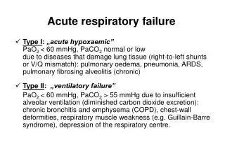
By Campbell (1965) Respiratory Failure • PaO2 <60, and/or • PaCO2 >50 Pathophysiology Acute Chronic Hypoxemic (Type I) Hypercapnic (Type II)
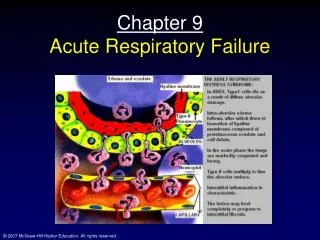
Physiology Normal respiration requires five separate components- • Nervous system- dorsal and ventral nuclei of respiratory control group, their afferent and efferent nuclei • Musculature- diaphragm, accessory msls and chest wall • Airways- up to the terminal bronchiole • Alveolar units • Vasculature
Intrapulmonary shunts • PaO2 falls progressively as shunt fraction increases but PaCO2 remaining constant unless shunt fraction exceeds 50% • The shunt fraction also determines the influence of inhaled oxygen on PaO2
Hypoxemic respiratory failure (type I) • V/Q mismatch • Shunt • Alveolar hypoventilation • Diffusion impairment • P/D impairment • FiO2 • Venous admixture
1. V/Q mismatch • Even in normal lungs areas present where V/Q mismatching is present ,ie, V/Q - at apex - at bases but overall a balance is maintained to achieve a steady ratio of ‘0.8’ for the whole lung (in healthy lungs) • In certain pathological states, the imbalance occurs leading to hypoxemia and V/Q mismatching
Pathophysiology • Obstruction • Fluid filled alveoli • Atelectasis, e.g. • Obstructive lung disease • Bronchospasm • Mucous plugging • Inflammation • Premature airway closure asthmatic exacerbation • Infection • CHF • Inhalational injury • ARDS
Clinical features • Hypoxia • Dyspnea, TC, tachypnea • Use of accessory muscle (nasal flaring) • Cyanosis – peripheral or central • Irritability • Confusion / coma Other signs – Specific to disease process • Radiologically V/Q mismatch • Black – hyperinflated lungs, e.g. COPD • White – in c/o occluded alveoli
Shunt • Extreme version of V/Q mismatch in which there is no ventilation to match perfusion (V/Q = 0) • Normal anatomical shunt • 2-3% • bronchial and thebesian veins Pathological anatomical shunt • R L blood flow through cardiac openings (e.g. ASD/VSD) or in pulm A-V malformations Physiological shunt • Atelectasis, pulmonary edema, or pneumonia does not generally respond to O2 supplementation as the gas exchange units are completely closed / collapsed (the alveolus)
2. Diffusion impairment Diffusion is movement of gas across the alveolar capillary membrane d/t pressure gradient. Any impairment in diffusion leads to inability of O2 to pass to the arterial blood, thereby lowering the arterial PaO2. e.g., • ILD, emphysema, pulm vascular abnormality, anemia, pulm HTN
Clinical features • Rarely – present as acute hypoxemia • ILD • Dry cough • Crackles (fine, basilar) • Clubbing of nailbeds • Rheumatological manifestations • Raynaud’s disease • S/o PHTN
3. Perfusion – diffusion impairment • Rare cause with liver disease complicated by hepatopulmonary syndrome • R L intracardiac shunts + Dilated pulm capillaries Thus, normal partial pressure of O2 are insufficient to drive O2 molecule through dilated pulm capillary Supplemental O2 Impaired gas exchange
4. ↓ PiO2 • barometric pressure (high altitude) 5. ↓ Mixed venous oxygen saturation The oxygen content in arterial blood represents the sum of oxygen in mixed venous blood and O2 added from alveolar air. • Normal gas exchange- PAO2 is major determinant of PaO2 • Impaired gas exchange- PvO2 determines PaO2 Thus a fall in mixed venous oxygen saturation can aggravate hypoxemia caused by V/Q abnormality.
Mixed venous oxygen saturation • PvO2 = k x DO2/VO2 DO2- delivery of oxygen to tissues VO2- uptake of oxygen by the tissues E.g., CHF with low cardiac output low Hb ↑O2 consumption
Hypercapnic respiratory failure (type II) (pump failure / ventilatory failure) Features • Elevated PaCO2 – creating an uncompensated respiratory acidosis • PaCO2 1/ alveolar ventilation Vd / VT CO2 production • Hypoxemia occurs d/t displacement of alveolar PO2 by CO2
Causes 1. Alveolar hypoventilation • ventilatory drive • Neurologic disease – impaired strength with failure of NM function in resp system 2.↑ Work of breathing 3. V/Q abnormality • Shunt – in late stages (edema, infiltrates) • Dead space ventilation- Vd/Vt > 50% (e.g., advanced emphysema ) 4. ↑ CO2 production , esp in patients with reduced ability to eliminate CO2.
Ventilatory drive • Chemoreceptor • Central (medullary) • Peripheral (aortic & carotid bodies) “Respond to CO2 & O2 tension” The drive can be diminished by • Drugs (overdose / sedation) • Brainstem lesions • Diseases of the CNS (multiple sclerosis, parkinsons) • Trauma to the brainstem • Hypothyroidism • Obesity • Sleep apnea • Metabolic alkalosis • Malnutrition • ICP • Metabolic encephalopathy
Neuromuscular disease CNS is stimulated but signal does not reach the goal • Reduced strength (d/t impaired NM transmission), e.g. • Spinal trauma • Motor neuron disease (amyotrophic lateral sclerosis or poliomyelitis) • MND (GBS) • NMJ disorder (MG/botulinism) • Respiratory muscle weakness • Muscular disease (muscle dystrophy, myositis, critical care myopathy, metabolic disorders ,hypophosphatemia, hypomagnesemia)
Work of breathing Pathophysiologically • Resistive load (bronchospasm) • Loads d/t lung compliance (alveolar edema, atelectasis, auto PEEP) • Loads d/t chest wall compliance (pneumothorax, PE, abd distension) • Load d/t MV requirement (PE, sepsis)
Clinically • airway resistance - COPD, asthma • Thoracic abnormalities – pneumothorax, rib #Ħ, pleural effusion, other RLD • CO2 production as in hypermetabolic state, e.g. – extensive burns
Type III resp failure • d/t lung atelectasis • Most commonly seen in periop period – perioperative resp failure • FRC collapse of dependent lung units Type IV resp failure • Due to hypoperfusion of respiratory muscles in shock (Harrison’s principles of internal medicine- 16th edition)
Diagnosis History e.g., • previous cardiac disease, VHD, recent symptom of orhopnea, chest pain • Trauma • Aspiration • Sepsis • Infection, etc
Physical examination Signs of • underlying disease process – pneumonia, pulmonary edema, asthma, COPD, corpulmonale etc • Hypoxemia- restlessness, anxiety, confusion, somnolence, tachycardia, dyspnea, cyanosis, use of accessory muscles, arrhythmias, seizures etc • Hypercapnia – asterixis, tachycardia, hypertension etc
Investigations • Disease specific- Hb, LFT, KFT, S. Electrolytes, Thyroid profile • ABG remains the confirmatory diagnostic tool- . PaO2 PaCO2 AaDO2 pH HCO3 O2 saturation • Radiological tools- CXR, CT- chest, V/Q scan, ECHO • Pulmonary function test- more useful in chronic resp failure
Treatment Admission to respiratory care unit or ICU • Hypoxemia • Hypercapnia • Underlying disease process
Guidelines for Standards of Care for Patients with Acute Respiratory Failure On Mechanical Ventilatory Support : Task Force on Guidelines Society of Critical Care Medicine Crit Care Med 1991 Feb; 19(2):275-278
Treatment • Airway management – ETT if required • Correction of hypoxemia- goal is to achieve a SaO2 of >90%, and a PaO2 of >60 mm Hg -supplemental oxygen - NIPPV - intubation and mechanical vent
Treatment • Correction of coexistent hypercapnia and respiratory acidosis • Ventilator management -non invasive -invasive
Non-invasive ventilatory support The application of ventilatory support through a nasal prong or full face mask in lieu of ETT is being used increasingly for patients with • acute or chronic mild to moderate respiratory failure • Conscious • Intact airway • Intact airway reflexes The various modes by which NIPPV can be provided include volume assist control, pressure assist control, BiPAP, CPAP, etc
Non-invasive ventilatory support Has proven beneficial in • acute exacerbations of COPD and asthma, • decompensated CHF with mild-to-moderate pulmonary edema, • pulmonary edema from hypervolemia • obesity hypoventilation syndrome Guidelines for noninvasive ventilation in acute respiratory failure. Indian J Crit Care Med 2006;10:117-47
Invasive ventilatory support • Useful when pt does not respond to non invasive methods of ventilation • Indications for IPPV based on specific threshold values for PCO2 and pH or arterial oxygenation have not been validated by clinical evidence
Invasive ventilatory support Indications- • Apnea or bradypnea • ALI & ARDS • Severe cardiogenic shock • Traumatic brain injury • Brain injury Indications for Invasive Mechanical Ventilation in Adults with Acute Respiratory FailureConference proceedings- Respiratory Care 2002, 47(3)
Non-invasive vs invasive ventilatory support • Guidelines for noninvasive ventilation in acute respiratory failure. Indian J Crit Care Med 2006;10:117-47 • Indications for Invasive Mechanical Ventilation in Adults with Acute Respiratory FailureConference proceedings- Respiratory Care 2002, 47(3)
Non-invasive vs invasive ventilatory support • NIV has been shown to be an effective treatment for acute hypercapnic respiratory failure (AHRF), particularly in chronic obstructive pulmonary disease (COPD). Facilities for NIV should be available 24 hours per day in all hospitals likely to admit such patients • NIV should not be used as a substitute for tracheal intubation and invasive ventilation when the latter is clearly more appropriate Non-invasive ventilation in acute respiratory failure. Thorax 2002;57:192–211 (British Thoracic Society Standards of Care Committee)
Follow up Complications • Pulmonary – embolism, barotrauma, fibrosis, cx secondary to use of mechanical devices, VILI, oxygen toxicity • Cardiovascular- hypotension, reduced cardiac output, arrhythmia, pericarditis, and acute myocardial infarction.
Gastrointestinal- hemorrhage, gastric distention, ileus, diarrhea, and pneumoperitoneum. • Infectious- pneumonia, urinary tract infections, and catheter-related sepsis • Renal - Acute renal failure and abnormalities of electrolytes and acid-base homeostasis • Nutritional – malnutrition, hypoglycemia, abd distension, etc
References • Miller’ s anaesthesia- 7th edition • Egans- Fundamentals of respiratory care-9th edition • The ICU Book- Paul Marino – 3rd edition • Harrison’s principles of internal medicine- 16th edition • Emedicine from WebMD: accessed from emedicine.com • Guidelines for noninvasive ventilation in acute respiratory failure. Indian J Crit Care Med 2006;10:117-47 • Indications for Invasive Mechanical Ventilation in Adults with Acute Respiratory FailureConference proceedings- Respiratory Care 2002, 47(3) • Non-invasive ventilation in acute respiratory failure Thorax 2002;57:192–211 (British Thoracic Society Standards of Care Committee) • Treatment of Acute Exacerbations of Chronic Respiratory Failure Chest 2004;125; 2217-2223 • Guidelines for Standards of Care for Patients with Acute Respiratory Failure On Mechanical Ventilatory Support :Crit Care Med 1991 Feb; 19(2):275-278