Download
1 / 54
800 likes | 2.15k Vues
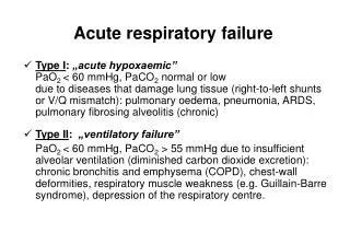
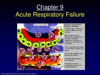
Acute respiratory failure. Classification of RF. Type 1 Hypoxemic RF ** PaO2 < 60 mmHg with normal or ↓ PaCO2 Associated with acute diseases of the lung Pulmonary edema (Cardiogenic, noncardiogenic (ARDS), pneumonia, pulmonary hemorrhage, and collapse. Type 2 Hypercapnic RF

E N D
Classification of RF • Type 1 • Hypoxemic RF ** • PaO2 < 60 mmHg with normal or ↓ PaCO2 • Associated with acute diseases of the lung • Pulmonary edema (Cardiogenic, noncardiogenic (ARDS), pneumonia, pulmonary hemorrhage, and collapse • Type 2 • Hypercapnic RF • PaCO2 > 50 mmHg • Hypoxemia is common • Drug overdose, neuromuscular disease, chest wall deformity, COPD, and Bronchial asthma
Distinction between Acute and Chronic RF • Acute RF • Develops over minutes to hours • ↓ pH quickly to <7.2 • Example; Pneumonia • Chronic RF • Develops over days • ↑ in HCO3 • ↓ pH slightly • Polycythemia, Corpulmonale • Example; COPD
More definitions • Hypoxemia = abnormally low PaO2 • Hypoxia = tissue oxygenation inadequate to meet metabolic needs • Hypercarbia = elevated PaCO2 • Respiratory failure may be acute or chronic
●Hypoventilation ●V/P mismatch ●Shunt ●Diffusion abnormality Pathophysiologic causes of Acute RF
CO2 O2
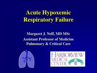
Mechanisms of hypoxemia • Alveolar hypoventilation • V/Q mismatch • Shunt • Diffusion limitation • Other issues we will not consider • Low FIO2 • Low barometric pressure
FIO2 Ventilation without perfusion (deadspace ventilation) Hypoventilation Diffusion abnormality Normal Perfusion without ventilation (shunting)
Perfusion without ventilation (shunting) Intra-pulmonary • Small airways occluded ( e.g asthma, chronic bronchitis) • Alveoli are filled with fluid ( e.gpulm edema, pneumonia) • Alveolar collapse ( e.g atelectasis)
Dead space ventilation • DSV increase: • Alveolar-capillary interface destroyed e.g emphysema • Blood flow is reduced e.g CHF, PE • Overdistended alveoli e.g positive- pressure ventilation
FIO2 Ventilation without perfusion (deadspace ventilation) Hypoventilation Diffusion abnormality Normal Perfusion without ventilation (shunting)
Hypercarbia • Hypercarbia is always a reflection of inadequate ventilation • PaCO2 is • directly related to CO2 production • Inversely related to alveolar ventilation PaCO2 = k x VCO2 VA
Hypercarbia • When CO2 production increases, ventilation increases rapidly to maintain normal PaCO2 • Alveolar ventilation is only a fraction of total ventilation VA = VE – VD • Increased deadspace or low V/Q areas may adversely effect CO2 removal • Normal response is to increase total ventilation to maintain appropriate alveolar ventilation
Common causes Hypoxemic RF typI HypercapnicRF typ II Chronic bronchitis,emphysema Severe asthma, drug overdose Poisonings, Myasthenia gravis Polyneuropathy, Poliomyelitis Primary ms disorders 1ry alveolar hypoventilation Obesity hypoventilation synd. Pulmonary edema, ARDS Myxedema, head and cervical cord injury Pneumonia, pulmonary edema Pulmonary embolism, ARDS Cyanotic congenital heart disease
Brainstem Spinal cord Airway Nerve root Nerve Lung Pleura Neuromuscular junction Chest wall Respiratory muscle Sites at which disease may cause ventilatory disturbance
1 – CNS Depression of the neural drive to breath Brain stem tumors or vascular abnormality Overdose of a narcotic, sedative Myxedema, chronic metabolic alkalosis Acute or chronic hypoventilation and hypercapnia Causes
Causes • 2 - Disorders of peripheral nervous system, Respiratory ms, and Chest wall • Inability to maintain a level of minute ventilation appropriate for the rate of CO2 production • Guillian-Barre syndrome, muscular dystrophy, myasthenia gravis, KS, morbid obesity • Hypoxemia and hypercapnia
Causes • 3 - Abnormities of the airways • Upper airways • Acute epiglotitis • Tracheal tumors • Lower airway • COPD, Asthma, cystic fibrosis • Acute and chronic hypercapnia
Causes • 4 - Abnormities of the alveoli • Diffuse alveolar filling • hypoxemic RF • Cardiogenic and noncardiogenic pulmonary edema • Aspiration pneumonia • Pulmonary hemorrhage • Associate with Intrapulmonary shunt and increase work of breathing
Hypoxemia Dyspnea, Cyanosis Confusion, somnolence, fits Tachycardia, arrhythmia Tachypnea (good sign) Use of accessory ms Nasal flaring Recession of intercostal ms Polycythemia Pulmonary HTN, Corpulmonale, Rt. HF Hypercapnia ↑Cerebral blood flow, and CSF Pressure Headache Asterixis Papilloedema Warm extremities, collapsing pulse Acidosis (respiratory, and metabolic) ↓pH, ↑ lactic acid Diagnosis of RF1 – Clinical (symptoms, signs)
Respiratory FailureSymptoms CNS: Headache Visual Disturbances Anxiety Confusion Memory Loss Weakness Decreased Functional Performance
Respiratory FailureSymptoms Pulmonary: Cough Chest pains Sputum production Stridor Dyspnea
Respiratory FailureSymptoms Cardiac: Orthopnea Peripheral edema Chest pain Other: Fever, Abdominal pain, Anemia, Bleeding
Clinical • Respiratory compensation • Sympathetic stimulation • Tissue hypoxia • Haemoglobin desaturation
Clinical • Respiratory compensation • Tachypnoea RR > 35 Breath /min • Accessory muscles • Recesssion • Nasal flaring • Sympathetic stimulation • Tissue hypoxia • Haemoglobin desaturation
Clinical • Respiratory compensation • Sympathetic stimulation • HR • BP • Sweating Tissue hypoxia • Altered mental state • HR and BP (late) • Haemoglobindesaturation cyanosis
Clinical Altered mental state ⇓PaO2 +⇑PaCO2 ⇨ acidosis ⇨ dilatation of cerebral resistance vesseles ⇨ ⇑ICP Disorientation Headache coma asterixis personality changes
Respiratory FailureLaboratory Testing Arterial blood gas PaO2 PaCO2 PH Chest imaging Chest x-ray CT sacn Ultrasound Ventilation–perfusion scan
Distinction between Noncardiogenic (ARDS) and Cardiogenic pulmonary edema ARDS Pulmonary edema
90 Hb saturation (%) 8 PaO2 (kPa) Pulse oximetry • Sources of error • Poor peripheral perfusion • Excessive motion • Carboxyhaemoglobin or methaemoglobin
Case 1 • A 36 yo man who has had a recent viral illness now is admitted to the ICU with rapidly progressive ascending paralysis (diagnosed as Guillain-Barre Syndrome). He is breathing shallowly at 36/min and complains of shortness of breath. His lungs are clear on exam. CXR shows small lung volumes without infiltrates. With the patient breathing room air, ABG are obtained. pH= 7.18 PaCO2= 68 mm Hg PaO2 =49 mm Hg HCO3=14mmol/l His hypoxemia is due to alveolar hypoventilation ACUTE RESP FALURE
Indications for intubation and mechanical ventilation • inability to protect the airway • respiratory acidosis (pH<7.2) • refractory hypoxemia • fatigue/increased metabolic demands • impending respiratory arrest • pulmonary toilet
Case 2 • A 65 yo man has smoked cigarettes for 50 yrs. He has chronic cough with sputum production and chronic dyspnea on exertion (stops once when climbing 1 flight of stairs). He is now admitted with several days of increased cough productive of green sputum and is short of breath even at rest. On exam his breathing is labored (32/min) and his breath sounds are quite distant. The expiratory phase is greatly prolonged and there are soft wheezes in expiration. pH=7.38 PCO2=48 PO2=48 O2 sat=78% HC03=38mmol/l His hypoxemia is predominantly due to V/Q mismatch chronic respiratory acidosis
Case 2- treatment • Supplemental oxygen • Nasal canula • Humidified mask • Venturi mask • Reservoir mask • Endotracheal tube • The goal of therapy is to achieve adequate oxygen content for O2 delivery.
Case 2 - treatment • The patient received 100% oxygen by reservoir mask and a small dose of medication to help him relax. • One hour later he is hard to arouse and his ABG shows pH 7.25, PaCO2 64, PaO2 310 • Has he improved? • What is his acid-base status now? • What happened?
Oxygen therapy • Like most other therapies, Oxygen therapy has both benefits and risks • Potential complications of oxygen therapy • Acute lung injury • Retrolental fibroplasia • Decreased respiratory drive in individuals with chronic hypercarbia • Use the lowest possible FIO2 to achieve adequate O2 saturation for oxygen delivery
Case 3 • A 56 yo man with known coronary artery disease and a prior myocardial infarction has had 1 hr of substernal chest pressure associated with nausea and diaphoresis. When you first see him, he is sitting upright in obvious distress and is cyanotic. He is breathing 36/min with short, shallow breaths. On examination of the chest he has dense inspiratory rales (crackles) half way up his back on both sides. Cardiac exam reveals faint heart sounds with an S3 gallop.
Case-3 ABG’s Mechanism of hypoxemia shunt CARDIOGEN PULMONARY EDEM
Respiratory physiology of congestive heart failure • Vascular congestion – increased capillary blood volume, mild bronchoconstriction, mild decrease in lung compliance; PaO2 normal or even increased • Interstitial edema – decreased compliance and lung volumes, worsening dyspnea, V/Q abnormality and widened A-a O2 gradient • Alveolar flooding – lung units that are perfused but not ventilated, shunt physiology with profound gas exchange abnormalities, decreased compliance and lung volumes
Treatment of cardiogenic pulmonary edema • Correct the problem with left ventricular function • Diruetics • Nitrates • Vasodilators • Thrombolytics, etc. • Decrease work of breathing • Ventilatory support • Improve oxygenation • Supplemental oxygen • Mechanical ventilation
ARDS Tachypnea, dyspnea, crackles Aspiration, sepsis 3 to 4 quadrant of alveolar flooding with normal heart size, systolic, diastolic function Decreased compliance Severe hypoxemia refractory to O2 therapy PCWP is normal <18 mm Hg Cardiogenic edema Tachypnea, dyspnea, crackles Lt ventricular dysfunction, valvular disease, IHD Cardiomegaly, vascular redistribution, pleural effusion, perihilar bat-wing distribution of infiltrate Hypoxemia improved on high flow O2 PCWP is High >18 mmHg Distinction between Noncardiogenic (ARDS) and Cardiogenic pulmonary edema
ICU admition 1 -Airway management Endotracheal intubation: Indications Severe Hypoxemia Altered mental status Importance precise O2 delivery to the lungs remove secretion ensures adequate ventilation Management of ARF
2 -Correction of hypoxemia O2 administration via nasal prongs, face mask, intubation and Mechanical ventilation Goal: Adequate O2 delivery to tissues PaO2 = > 60 mmHg Arterial O2 saturation >90% Management of ARF
4 – Mechanical ventilation Indications Persistence hypoxemia despite O2supply Decreased level of consciousness Hypercapnia with severe acidosis (pH< 7.2) Management of ARF
4 - Mechanical ventilation Increase PaO2 Lower PaCO2 Rest respiratory ms (respiratory ms fatigue) Ventilator Assists or controls the patient breathing The lowest FIO2 that produces SaO2 >90% and PO2 >60 mmHg should be given to avoid O2 toxicity Management of ARF
5 -PEEP (positive End-Expiratory pressure Used with mechanical ventilation Increase intrathoracic pressure Keeps the alveoli open Decrease shunting Improve gas exchange Hypoxemic RF (type 1) ARDS Pneumonias Management of ARF
6 - Noninvasive Ventilatory support (IPPV) Mild to moderate RF Patient should have Intact airway, Alert, normal airway protective reflexes Nasal or full face mask Improve oxygenation, Reduce work of breathing Increase cardiac output AECOPD, asthma, CHF Management of ARF
7 - Treatment of the underlying causes After correction of hypoxemia, hemodynamic stability Antibiotics Pneumonia Infection Bronchodilators (COPD, BA) Salbutamol reduce bronchospasm airway resistance Management of ARF