Human Eye
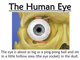
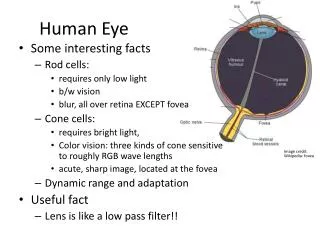
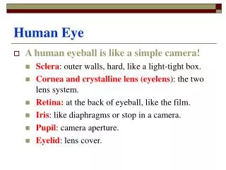
Human Eye. A human eyeball is like a simple camera! Sclera : outer walls, hard, like a light-tight box. Cornea and crystalline lens (eyelens ): the two lens system. Retina: at the back of eyeball, like the film. Iris : like diaphragms or stop in a camera. Pupil : camera aperture.

Human Eye
E N D
Presentation Transcript
Human Eye • A human eyeball is like a simple camera! • Sclera: outer walls, hard, like a light-tight box. • Cornea and crystalline lens (eyelens): the two lens system. • Retina: at the back of eyeball, like the film. • Iris: like diaphragms or stop in a camera. • Pupil: camera aperture. • Eyelid: lens cover.
Sclera (The white/non-transparent tissue surrounding the cornea)
Aqueous humor and Vitreous humor • The Aqueous Humor is the clear liquid between the cornea and the lens. It has the benefit of being fairly homogenous and, as a result, the optical properties are easily measured. (Le Grand, 1967) • The space that it inhabits is called the anterior chamber. • The Vitreous Humor is the clear liquid between the lens and the retina. • The space that it fills is called the vitreous body.
Functions? • Provides nourishment to the eyelens and cornea. Cannot use the blood vessels: • Will block the light. • Easy for surgical transplant. • Hold the shape of the eyeball.
Focusing • The cornea and eyelens form a compound lens system, producing a realinverted image on the retina. • From air to cornea (n=1.376): large bending, the main focusing. • From cornea to eyelens (n=1.406), less focusing power. (Eyelens can develop white cloudiness when getting old: Cataracts.) • The eye has a limited depth of field. We cannot see things close and far at the same time.
Accommodation • The eye focusing is not done by change the distance between the lens and retina. Rather, it is done by changing the focal length of the eyelens! Ciliary muscles help to change the shape of the lens: accommodation. • Muscles relax, long focal length, see objects far way; Muscles tense, short focal length see objects close. • Normal eyes can see 25cm to infinity, however, if the cornea bulges too much or too little. The accommodation does not help. (myopia or hyperopia)
The Iris • When it is full open, it is about f/2 and f/3. This happens at low light level. • When the iris has a small opening, it can cut down the light intensity by a factor of 20. • However, the main function of stopping down the iris is to increase the depth of field.
Retina Structure • Light sensitive layer is made of photo-receptors: rods (120 millions) and cones (7 millions) which absorb the light. • Plexiform Layer: nerve cells that process the signals generated by rods and cones and relay them to the optical nerve. • Choroid: carries mayor blood vessels to nourish the retina and absorb the light so that it will not be reflected back (dark pupil!)
Rods and Cones • Covers an area of 5 cm2. A baseball a mile away gives an image covering one cone. • Cones: for more precise vision, need strong light. help to see colors. Mostly distributed in the center of the retina (fovea). • Rods: for peripheral and night vision. Sensitive to light. Mostly distributed away from fovea.
Sensitivity • Cones: slow, fine grain, like color film. • Need high level of light (photopic condition, day) • High density, high resolution. • Rods: fast, coarse grain, black & white film • Low level of light (scotopic condition, at night) No color is obvious. • Adaptation: Changing of retina sensitivity.
Singal Processing • Trace the signal through the retina: • The retina is a seven-layered structure involved in signal transduction. • Light enters from the GCL side first, and must penetrate all cell types before reaching the rods and cones. • The outer segments of the rods and cones transduce the light and send the signal through the cell bodies of the ONL and out to their axons.
In the OPL photoreceptor axons contact the dendrites of bipolar cells and horizontal cells. Horizontal cells are interneurons which aid in signal processing • The bipolar cells in the INL process input from photoreceptors and horizontal cells, and transmit the signal to their axons.
In the IPL, bipolar axons contact ganglion cell dendrites and amacrine cells, another class of interneurons. • The ganglion cells of the GCL send their axons through the OFL to the optic disk to make up the optic nerve. They travel all the way to the lateral geniculate nucleus.
Fovea • The fovea defines the center of the retina, and is the region of highest visual acuity. The fovea is directed towards whatever object you wish to study most closely - this sentence, at the moment. In the fovea there are almost exclusively cones, and they are at their highest density.
Processing Time • Latency: it takes a bit time for the cells in retina to respond to a flash of light. • Persistence of response: the response does not stop at the instant the flash stops. • 1/25 second at low intensity, 1/50 second at high intensity. • The persistence allows as to see moving things clearly.