Neuron
ud83eudde0 Types of NeuronsnSensory NeuronsnCarry information from sense organs to the brain and spinal cord.nMotor NeuronsnTransmit signals from the brain/spinal cord to muscles and glands.nInterneuronsnConnect neurons within the brain and spinal cord.


Neuron
E N D
Presentation Transcript
Israr Hussain YousafzaiBS (Anesthesia) CRT PGDRT MSPHSenior Respiratory Therapist, HMC Peshawar Neuron
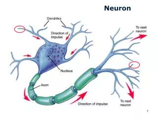
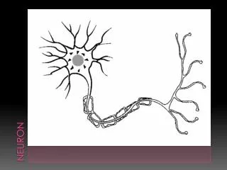
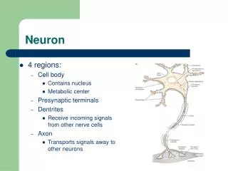
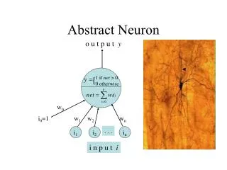
Neuron • Neuron or nerve cell is the structural and functional unit of nervous system • Neuron is similar to any other cell in the body, having nucleus and all the organelles in cytoplasm • However, it is different from other cells by two ways: • Neuron has branches or processes called axon and dendrites (nerve fiber) • Neuron does not have centrosome. So, it cannot undergo division
Classification of Neuron • Neurons are classified by three different methods: • Depending upon the number of poles • Depending upon the function • Depending upon the length of axon
Classification of Neuron Cont’d... 1. Depending Upon The Number Of Poles • Pole is the point from which the nerve fibers arise • Based on the number of poles, neurons are divided into three types: • Unipolar neurons – embryonic stage • Bipolar neurons • Multipolar neurons
Classification of Neuron Cont’d... 2. Depending Upon The Function • On the basis of function, nerve cells are classified into two types: • Motor or efferent neurons • Long axon and short dendrites • Carry information from CNS to periphery • Sensory or afferent neurons • Short axon and long dendrites • Carry information from periphery to CNS
Classification Of Neuron Cont’d... 3. Depending Upon The Length Of Axon • Depending upon the length of axon, neurons are divided into two types: • Golgi type I neurons • Cell body is in different parts of CNS while long axon extends to effectors • Golgi type II neurons • Short axons, found in cerebral cortex and spinal cord
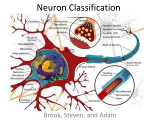
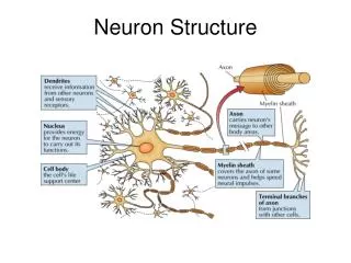
Structure Of Neuron • Neuron is made up of three parts: • Nerve cell body • Dendrite • Axon • Dendrite and axon form the processes of neuron • Dendrites and axons are usually called nerve fibers
Nerve Cell Body • Soma or perikaryon • It is irregular in shape • Neuro membrane • Neuroplasm: • Large nucleus • Nissl’s bodies • Neurofibrils • Other same as usual cell
Nerve Cell Body • Nucleus • Single and large • One or two prominent nucleoli • Does not contain centrosome Neurofibrils • Thread like structures present in the form of network • Consist of microfilaments and microtubules • Present in soma and the nerve processes
Nerve Cell Body Nissl’s Bodies • Basophilic – purple • Tigroid substances (spotted) • Containing ribosomes • Found in cell body and dendrites, not in axon • Distinguishing dendrites from axons
Axon • Each neuron has only one axon • Axon arises from axon hillock of the nerve cell body having no Nissl’s granules • Length of longest axon is about 1 meter • Axon transmits impulses away from the nerve cell body
Axon • Axis Cylinder • Axon has a long central core of cytoplasm called axoplasm • Axoplasm is covered by tubular sheath likemembrane called axolemma • Axolemma is the continuation of the cell membrane of nerve cell body • Axoplasm contains mitochondria, neurofibrils and axoplasmic vesicles • Axoplasm along with axolemma is called the axis cylinder
Axon • As Nissl’s bodies are absent in the axon, necessary proteins are synthesized in the soma, and are transported from soma to axon, by means of axonal flow • Axis cylinder of the nerve fiber is covered by a membrane called neurilemma
Dendrites • Branched process of neuron that branched repeatedly • It may be present or absent • If present, it may be one or many in number • Dendrite has Nissl granules and neurofibrils • Dendrite transmits impulses towards the nerve cell body • Usually, the dendrite is shorter than axon
Organization Of Nerve • Fasciculus • Epineurium • Perineurium • Endoneurium
Myelin Sheath • Thick, lipoprotein sheath • it is not a continuous sheath, absent at regular intervals • The area where it is absent, is called node of Ranvier • Segment of nerve fiber b/w two nodes is called internode • It is formed by Schwann cells
Myelin Sheath • Myelinogenesis starts at 4th month of pregnancy and completed at 2ndyear after birth • Myelin sheath is responsible for the following: • White color of nerve fibers • Insulation • Faster and saltatory conduction
Neurilemma • Also called neurilemmal sheath or sheath of Schwann • It contains Schwann cells • In non-myelinated nerve fiber, it surrounds axolemma continuously and serves as a covering membrane • In myelinated nerve fiber, it covers the myelin sheathand are necessary for myelinogenesis
Neurilemma • At node of Ranvier, it invaginates and runs up to axolemma • Neurilemma is absent in CNS • So, the oligodendroglia are responsible for myelinogenesis
Neurotrophins – Neurotrophic Factor • The protein substances, essential for growth and functioning of nervous tissue • Secreted by many tissues in the body i.e. muscles, astrocytes and neurons Function • Facilitate initial growth and development of nerve cells in CNS and PNS • Promote survival and repair of the nerve cells • Maintenance of nervous tissue and neural transmission Examples NGF, BDGF, CNTF, GDNF, FGF, NT-3
Classification Of Nerve Fibers • 1. Depending upon structure – myelinated, non myelinated • 2. Depending upon distribution – somatic, autonomic • 3. Depending upon origin– cranial, spinal • 4. Depending uponfunction – sensory, motor • 5. Depending upon secretion of neurotransmitter– adrenergic, cholinergic • 6. Depending upon diameter and conduction of impulse • (Erlanger Gasser classification)
Erlanger Gasser’s Classification • Type A nerve fibers (alpha, beta, gamma and delta) • Type B nerve fibers • Type C nerve fibers • Except type C fibers, all the nerve fibers are myelinated • Thickness is directly proportional to velocity of conduction