Download
1 / 22
230 likes | 402 Vues
Receptors & Signaling. Assumed Knowledge . Structure of membrane proteins Ion concentrations across membranes Second messengers in signal transduction Regulation of protein activity through phosphorylation. Membrane Proteins . Mainly transmembrane Act as receptors

E N D
Assumed Knowledge • Structure of membrane proteins • Ion concentrations across membranes • Second messengers in signal transduction • Regulation of protein activity through phosphorylation
Membrane Proteins • Mainly transmembrane • Act as receptors • Ligand binding causes a conformational change – a change in the shape of the receptor
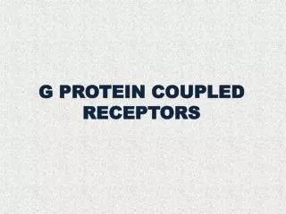
G Protein coupled receptors (GPCR) – these have a transmembrane bit with 7 helices spanning the membrane The extracellular part binds to the ligand The intracellular part binds to the G protein
G Protein – these are proteins that bind to the guanine nucleotide (GTP – guanosine triphosphate, GDP – guanosine diphosphate) Hydrolysis of GTP releases a phosphate group which can act on other molecules – transmits the signal GTP > GDP + P
G proteins have three subunits – α (alpha), β (beta), and γ (gamma). β and γsubunits are tightly bound together. α binds to GDP
Ligand binding to the transmembrane protein causes a conformational change and release of the α subunit The α subunit exchanges GDP > GTP and becomes active The α subunit meets a target and phosphorylates it (adds a phosphate group from GTP converting it to GDP – this is hydrolysis of GTP) Hydrolysis = cleavage Phosphorylation = addition of a phosphate group
Now the α subunit is bound to GDP, it becomes inactive again and re-associates with the transmembrane protein and the β and γ subunits
Ion Concetrations Ions – these are molecules with a charge + or – Ion concentration – ions move down their concentrations gradient from [high] to [low]
- + - + - + - + - + - + - + - + Membrane potential = -70 mV Anion –ve charge Cation +ve charge [K+] 160 mM [K+] 5 mM [A-] 165 mM [A-] 40 mM [Na+] 150 mM [Na+] 10 mM [Cl-] 115 mM [Cl-] 5 mM [Ca2+] 2 mM [Ca2+] 0.2 M
Membrane Potential – this is the difference in charge between the interior and exterior of a cell Typically the interior is more negative
Action potentials – these are rapid rises and falls in the membrane potential. In neurons these act as nerve signals The interior becomes rapidly positive or less negative – depolarization. This is followed by a rapid return to a negative membrane potential – repolarization There is a transient hyperpolarization where the membrane potential becomes more negative than normal
1. An stimulus is received by a nerve causing Na+ channels to open – Na+ moves into the cell. The membrane potential begins to become more positive
When the membrane potential reaches the threshold level (-55mV) there is an opening of more Na+ channels allowing more Na+ to enter the cell This is an ‘all-or-nothing’ moment meaning if the membrane potential doesn’t reach the threshold there will be no action potential but if it reaches the threshold there no turning back
3. As Na+ enters the cell there is delayed opening of K+ channels The membrane potential reaches +30mV where the Na+ channels close
4. When the K+ open, K+ leaves the cell causing the membrane to start to become more negative again
When the K+ channels finally close there is slightly more K+ on the outside than Na+ meaning the membrane potential dips below the normal resting potential (-70mV) Essentially more positive charges leave the cell than entered
5. There is a refractory period where the [K+] and [Na+] are returned to their original state Carried out by the Na+/K+ -ATPase (a pump that uses ATP for energy)
Second Messengers Second messengers – theseare molecules that relay signals from receptors on the cell surface to target molecules inside the cell Examples include IP3, Ca2+, cAMP Allows for amplification of the signal