Download

1 / 93
950 likes | 1.42k Vues
NEUROANATOMY Lecture : 12 Anatomy of the Autonomic Nervous System (Involuntary Nervous System). Prepared and presented by: Dr. Iyad Mousa Hussein, MD, Ph.D of Neurology Head of Neurology Department Nasser Hospital. LECTURE OBJECTIVES:. Structures of the Autonomic Nervous System.

E N D
NEUROANATOMY Lecture : 12 Anatomy of the Autonomic Nervous System (Involuntary Nervous System) Prepared and presented by: Dr. Iyad Mousa Hussein, MD, Ph.D of Neurology Head of Neurology Department Nasser Hospital
LECTURE OBJECTIVES: Structures of the Autonomic Nervous System. Effects of Autonomic Nervous System on Organs and Body. The Origin of the Autonomic Nervous System. The Sympathetic Nervous System. The Cervical Part of the Sympathetic Chain. The Parasympathetic Nervous System. The Parasympathetic Ganglia in the Head and Neck. Type, Site, Roots, and Branches of the Submandibular Ganglion. Type, Site, Roots, and Branches of the Sphenopalatine Ganglion. Type, Site, Roots, and Branches of the Otic Ganglion. Type, Site, Roots, and Branches of the Ciliary Ganglion. Sympathetic Supply of the Pupil.
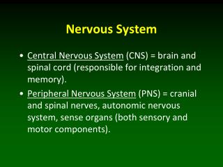
Anatomy of the Autonomic Nervous System (Involuntary Nervous System) It is part of the nervous system which supplies the viscera (glands, smooth and cardiac muscles).
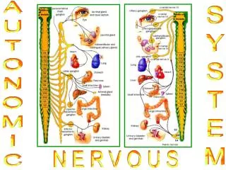
Structures of the Autonomic Nervous System • Sympathetic nervous system (Thoraco-lumbar). • Parasympathetic nervous system (Cranio-sacral).
How are the Visceral Structures Supplied by Autonomic Fibers? • The motor (autonomic) supply to the visceral structures in two points: • Most of the visceral structures have two nerve supply: one sympathetic and one parasympathetic. One of these nerves acts as a "stimulator" while their acts as an "inhibitor". • Each of two motor nerves (sympathetic and parasympathetic) is formed of a chain of two neurons: • a. The body of the first neuron is found in the CNS and • its axon is called a preganglionic fiber. • b. The body of the second neuron is found in the PNS (either in sympathetic or in parasympathetic ganglia) and its axon is called a postganglionic fiber.
The Origin of the Autonomic Nervous System • 1. The Origin of the Sympathetic Nervous System: • Consists of fibers which arise from the 12 thoracic and upper 2 lumbar segments (Thoraco-lumbar part) and reach the sympathetic chain by means of the white rami. • 2. The Origin of the Parasympathetic Nervous System: • a. The cranial part: consists of fibers which pass in • four cranial nerves (3, 7, 9 & 10 cranial nerves). • b. The sacral part: consists of fibers which arise • from the second, third and fourth sacral segments • of the spinal cord.
Transmitters of the Autonomic Nervous System 1. The preganglionic transmitter: For sympathetic and parasympathetic preganglionic transmitter is Acetylcholine. 2. The postganglionic transmitter: a. Parasympathetic postganglionic transmitter is Acetylcholine. b. Sympathetic postganglionic transmitter is Norepinephrine.
The Sympathetic Nervous System • The sympathetic NS consists of fibers which arise from the 12 thoracic and upper 2 lumbar segments (Thoraco-lumbar part) and reach the sympathetic chain by means of the white rami. • The Sympathetic Chain: • There are two sympathetic chains (right and left) on • either side of the vertebral column. • Each chain extends from the level of the first cervical • vertebra (above) to front of the coccyx (below) where • two chains unite to form a single coccygeal ganglion • (impar ganglion).
How Many Sympathetic Ganglia are Found in the Sympathetic Chains? Cervical:3 pairs. Thoracic:10-12 pairs. Lumbar:4-5 pairs. Sacral:4-5 pairs. Coccygeal:single coccygeal ganglion.
The Cervical Part of the Sympathetic Chain • Site: It lies behind the common, external, and the internal carotid arteries, outside carotid sheath and medial to the vagus nerve. • It has: • Preganglionic fibers:from upper thoracic segments until the three cervical ganglion. • Postganglionic fibers:leave these ganglia, to the cervical nerves and to cranial nerves.
The Cervical Sympathetic Ganglion • There are three cervical sympathetic ganglion: • Superior cervical sympathetic ganglion. • Middle cervical sympathetic ganglion. • Inferior cervical sympathetic ganglion.
The Superior Cervical Sympathetic Ganglion • It is the largest cervical sympathetic ganglion (about 2-3 cm long). • Site: lies behind the internal carotid artery. Its continuation upwards inside the skull is called the internal carotid nerve. • Branches: • Grey rami: to upper four cervical nerves. • Communicating branches: to the cranial nerves. • The internal carotid nerve: passes upwards to form the internal carotid plexus around the internal carotid artery. • The external carotid nerve: passes to the external carotid artery to form an external carotid plexus. • The pharyngeal branch: to form part of the pharyngeal plexus. • The cervical cardiac sympathetic branch: to the heart.
The Middle Cervical Sympathetic Ganglion • Site: lies on the inferior thyroid artery, behind the common carotid artery at the level C6 vertebra • Branches: • Grey rami: to the 5th and 6th cervical nerves. • The middle cervical cardiac sympathetic branch: to heart. • Thyroid branches: to the thyroid gland.
The Inferior Cervical Sympathetic Ganglion • Site: lies behind the common carotid artery and the lower part of the vertebral artery at the level C7 vertebra. • Branches: • Grey rami: to 7th and 8th cervical nerves. • The inferior cervical cardiac sympathetic branch: • to heart. • 3. Vertebral plexus: around the vertebral artery.
The Parasympathetic Nervous System • The parasympathetic NS consists of two parts: • The cranial part: consists of fibers which pass in four cranial nerves (3, 7, 9 & 10 cranial nerves). • The sacral part: consists of fibers which arise from the second, third and fourth sacral segments of the spinal cord.
The Parasympathetic Ganglia in the Head and Neck • There are four parasympathetic ganglia: • Submandibular ganglion. • Sphenopalatine ganglion. • Otic ganglion. • Ciliary ganglion.
Submandibular Ganglion • Type: it is a small parasympathetic ganglion (2-3 mm in diameter). • Site: it lies in the submandibular region on the upper part of hyoglossus muscle. • Relations: • Superiorly: lingual nerve. • Inferiorly: deep part of submandibular gland and its duct. • Medially: hyoglossus muscle. • Laterally: mylohyoid muscle.
Submandibular Ganglion • Roots Entering the Submandibular Ganglion: • Parasympathetic root: arising from the chorda tympani nerve. It joins the lingual nerve then the parasympathetic fibers leave lingual nerve to relay in the ganglion. • Sympathetic root: arising from the plexus around facial artery (passes without relay in the ganglion). • Sensory root: from lingual nerve (passes without relay in the ganglion). • Branches of the Submandibular Ganglion: • Several small branches (mixed: parasympathetic + sympathetic + sensory fibers) pass directly from the ganglion to supply the submandibular salivary gland. • Similar branches join the lingual nerve to reach the sublingual salivary gland.


![AUTONOMIC NERVOUS SYSTEM [ANS] DISCUSSION](https://cdn1.slideserve.com/2214545/autonomic-nervous-system-ans-discussion-dt.jpg)