Sickle Cell Disease
Sickle Cell Disease. Sickle Cell Anemia. Sickle Cell anemia is an inherited red blood cell disorder. Normal red blood cells are biconcave discs, and they move through small blood tubes in the body to deliver oxygen.


Sickle Cell Disease
E N D
Presentation Transcript
Sickle Cell Anemia Sickle Cell anemia is an inheritedred blood cell disorder. Normal red blood cells are biconcave discs, and they move through small blood tubes in the body to deliver oxygen. Sickle red blood cells become hard, sticky and shaped like sickles. When these hard and pointed red cells go through the small capillaries, they clog the flow and break apart. This can cause pain, damage and a low blood count, or anemia.
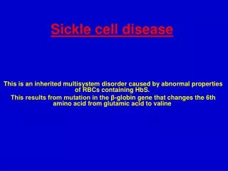
The origin of the disease is a small change in the protein hemoglobin A single change in an amino acid causes hemoglobin to aggregate. Glutamic acid is replaced by Valine in position 6 in the beta chain.
The replacement of valine for Glutamic acid produce an abnormal Hb called : HbS : (2α,2βs) which is insoluble and forms crystals when exposed to low O2 tension : polymerizes into long fibers.
Symptoms of anemia • Abdominal and bone/joint pain • Delayed growth and puberty • Jaundice • Greater risk for infection • Adolescents and adults can develop ulcers on their legs • Chest pain • Excessive thirst • Dactylitis About 30% of Jamaican patients with Sickle Cell develop ulcers in comparison to 1% of Americans Symptoms
Sickle cell is an autosomal recessive disease. • Therefore, the child can only get Sickle cell if both parents are carriers, not if only one is and the other is normal. They have a 25% chance of getting it if both are carriers Genetics of Sickle Cell
Three common types of Sickle Cell Disorders • Sickle Cell Anemia (Homozygous) • Hb SS • More common. • Severe hemolytic anemia. • Leg ulcers due to vascular stasis الركود الدموي& ischemia. • Most Severe – No HbA • Crises : (painful vaso-occlusive, visceral, a plastic or hemolytic) • Factors that cause crises include : infection, acidosis, dehydration, deoxygenation, cold, exercise)
2. Double heterozygous : (Hb SC, Hb S βthal.) • Sickle cell trait (Hb AS) • Benign • No anemia • RBC remain flexible with normal morphology • Hematuria is the most common symptom • The person is a Carrier • Hb S is : 25 – 45% • Care must be taken with anesthesia, pregnancy and high altitudes.
In Hb SS : Hb is between 6 – 9 g/dl. • The blood film show : sickle cells, target cells, Howell-Jolly bodies. • Positive Sickling test. • Hb electrophoresis : Hb SS : 80 – 100% no Hb A Hb F : 5 – 15% Lab. Findings
Blood film in sickle cell anemia : sickle cells, target cells, polychromatic cells
These are basophilic nuclear remnants (clusters of DNA) in circulating erythrocytes. • They are usually observed singly in hemolytic anemia, following splenectomy, and in cases of splenic atrophy. Howell-Jolly bodies
This is a wet preparation. • 5 drops of reagent (Sodium dithionite), are added to 1 drop of anticoagulated blood on a slide. Cover glass is put on and sealed with petrollium jelly/parraffin wax mixture. • The reagent is a reducing agent. • In Hb SS, sickling occur immediately, while it may take 1 hour in Hb S trait. The Sickling Test
Differentiates the various types of hemoglobins in the blood. • Electrophoresis, a process that causes movement of particles in an electric field, resulting in formation of "bands" that separate toward one end or the other in the field. • With the Cellulose Acetate Electrophoresisat Alkaline pH, Hb is a negatively charged protein and when subjected to electrophoresis will migrate toward the anode (+). Hb. electrophoresis
This is done after the Hb electrophoresis to differentiate between some hemoglobins that have the same electrophoretic mobility as HbS. (Hb D & Hb G) • Only Hb S precipitate in the reduced state when placed in a high molarity phosphate buffer. • 0.05 ml of blood is added to 1 ml of the buffer and mixed in a test tube. • Positive results : presence of Hb S : cloudy solution . • Negative results : other Hbs : clear solution . Hb S solubility Test