Download
1 / 27
270 likes | 428 Vues
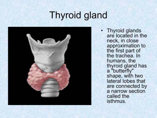
Digestive gland. The extrinsic glands of the digestive system include the major salivary glands, the pancreas, and the liver, all of which are located outside the wall of the digestive tract.

E N D
The extrinsic glands of the digestive system include the major salivary glands, the pancreas, and the liver, all of which are located outside the wall of the digestive tract. The major salivary glands consist of three pair of glands: the parotid, submandibular glands and sublingual glands.
Interlobular septum parenchyma Fig. 1 Submandibular gland : mixed gland
① serous demilune ② Fig. 2 Submandibular gland(HE stain,high mag.) ①Mixed acinus②Serous acinus
① Intercalated duct ② Fig.3 Submandibular gland(HE stain,high mag.) ①mucous acinus ②Serous acinus
① ② Fig.4 Submandibular gland(HE stain,high mag.) ①Secretory duct ②Serous acinus
Intralobular duct Fig.5 Submandibular gland (HE stain,high mag.)
The pancreas can be divided into two portion: exocrine portion and endocrine portion. The exocrine portion is a serous, compound tubloacinar gland. The endocrine portion is called pancreatic islet( islet of Langerhans).
② ① ① Fig. 6 Pancreas (HE stain, low mag.) ①pancreatic islet ②exocrine portion
Fig .7 Pancreas (HE stain, high mag.) centroacinar cells
① ② Fig. 8 Pancreas(HE stain, high mag.) ①pancreatic islet ②exocrine portion
② ① Fig . 9 A cell of pancreatic islet (TEM) ①nucleus ②secretory granule The cells secrete glucagon.
① ② Fig. 10 B-cell of pancreas islet(TEM)①nucleus ②secretory granule The B cells secrete insulin.
The liver is the biggest gland in the body. It can be divided into two main parts: hepatic lobule and portal area. The hepatic lobule includes five components :central vein, hepatic plate(cord), hepatic sinusoid, perisinusoidal space and bile canaliculi.
Hepatic lobule Portal areas Fig. 11 Liver of pig (HE stain, low mag.)
Hepatic sinusoid Hepatic cord Central vein Fig. 12 hepatic lobule (HE stain, high mag.)
② ③ ① Fig. 13 Portal area of liver(HE stain , high mag.) ①interlobular artery ②interlobular vein ③ interlobular bile duct
Central vein Central vein Fig. 14 Liver of human (HE stain, low mag.)
Hepatic macrophage Hepatic macrophage Fig. 15 Liver of mouse (trypan blue injected preparation, high mag.)
Fig. 16 Hepatic macrophage in hepatic sinusoid (SEM)
bile canaliculus Central vein Fig. 17 liver(silver stain, low mag.) showing bile canaliculus
bile canaliculus Fig. 18 liver (silver stain, high mag.) showing bile canaliculus
Three layers : mucous membrane, muscularis and serosa. plica Fig.19 gall bladder (HE, low mag.)
Simple columnar epithelium Fig.20 gall bladder (HE, low mag.)