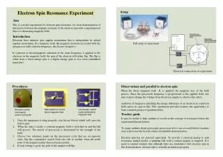
Download
1 / 27
400 likes | 1.27k Vues
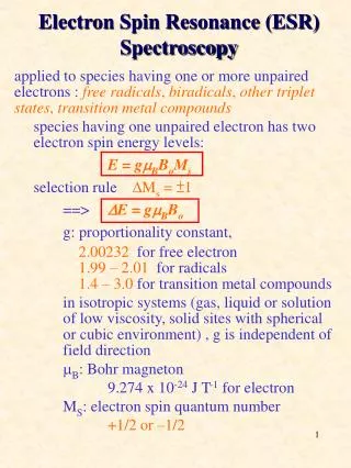
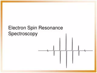
Electron Spin Resonance Spectroscopy. or It’s fun to flip electrons!. E lectron P aramagnetic R esonance spectroscopy. E lectron S pin R esonance spectroscopy. Classical theory: Electron spin moment interacts with applied electromagnetic radiation. D. E. B. 0. n. h.

E N D
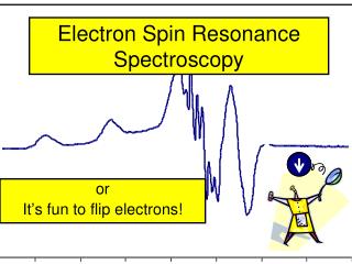
Electron Spin Resonance Spectroscopy or It’s fun to flip electrons!
Electron Paramagnetic Resonance spectroscopy Electron Spin Resonance spectroscopy
Classical theory: Electron spin moment interacts with applied electromagnetic radiation D E B 0 n h Principles of EMR spectroscopy Quantum theory: transitions between energy levelsinduced by magnetic field Resonance condition hn= gmBB0
The EPR experiment • Put sample into experimental magnetic field (B) • Irradiate (microwave frequencies) • Measure absorbance of radiation as f(B) Weil, Bolton, and Wertz, 1994, “Electron Paramagnetic Resonance”
The hyperfine effect • The magnetic field experienced by the unpaired electron is affected by nearby nuclei with non-zero nuclear spin Weil, Bolton, and Wertz, 1994, “Electron Paramagnetic Resonance”, New York: Wiley Interscience.
Hyperfine splitting of EPR spectra • The magnitude of the splitting and the number of lines depend upon: • The nuclear spin of the interacting nucleus • # of lines = 2n(I + ½) so I = ½ gives 2 lines, etc. • The nuclear gyromagnetic ratio • The magnitude of the interaction between the electronic spin and the nuclear spin • Magnitude of the splitting typically decreases greatly with increasing numbers of bonds between the nucleus and unpaired electron
No hyperfine Hyperfine coupling If the electron is surrounded by n spin-active nuclei with a spin quantum number of I, then a (2nI+1) line pattern will be observed in a similar way to NMR. In the case of the hydrogen atom (I= ½), this would be 2(1)(½) + 1 = 2 lines. 1 H) 14 N) 2 identical I=1/2 nuclei 17 1 I=5/2 nucleus ( O) 10 Gauss
Some nuclei with spins Element Isotope Nuclear No of % spin lines abundance Hydrogen 1H ½ 2 99.985 Nitrogen 14N 1 3 99.63 15N ½ 2 0.37 Vanadium 51V 7/2 8 99.76 Manganese 55Mn 5/2 6 100 Iron 57Fe ½ 2 2.19 Cobalt 59Co 7/2 8 100 Nickel 61Ni 3/2 4 1.134 Copper 63Cu 3/2 4 69.1 65Cu 3/2 4 30.9 Molybdenum 95Mo 5/2 6 15.7 97Mo 5/2 6 9.46
Hyperfine splittings multiply with the number of nuclear spins Benzoquinone anion radical: 1 proton – splits into 2 lines 1:1 2 protons split into 3 lines 1:2:1 3 protons split into 4 lines 1:3:3:1 4 protons split into 5 lines 1:4:6:4:1 -60 C At higher temperature: faster motion - sharper lines shorter lifetime - smaller signal 20 C
Prushan Example 77 K Cryogenic ESR Spectrum of [Cu(Thyclops)]ClO4 in MeOH Prushan, M. J.; Addison, A. W.; Butcher, R. J.; Thompson, L. K. “Copper(II) Complex Tetradentate Thioether-Oxime Ligands” Inorganica Chimica Acta, 358, 3449-3456 (2005).
2nI+1 2x2x1+1
Diagram of an ESR spectrometer Circulator Detector Klystron Microwave source Cavity N S cryostat Spectrophotometer Detector Light source
Hyperfine Splitting If the odd, unpaired electron is associated with a nucleus with nuclear spin, can get coupling between the two spins and observe 2I+1 (I = nuclear spin) “peaks” or “valleys”. Examples: di-t-butyl nitroxide radical; I(N) = 1;
Hyperfine Splitting vanadyl [V=O]2+ complex; I (V) = 7/2; 2(7/2) + 1 = 8 peaks
Signal Intensities Follow Pascal's triangle
superhyperfine splitting carbon compound; I(C) = 0; 2(0) + 1 = 1 peak…. But: If the odd, unpaired electron spends time around multiple sets of equivalent nuclei, additional splitting is observed: 2nI + 1; this is called “superhyperfine splitting.” Examples: Triplet Quartet Pentet
Superhyperfine Splitting Examples: Septet Sextet Octet Superhyperfine splitting is direct evidence for COVALENCY!
It is possible for the unpaired electron to spend differing amounts of time on different nuclei. The greater the covalency, the greater is the hyperfine splitting. Triplet: hyperfine splitting. Doublet: superhyperfine splitting. Interpretation: electron is spending most of its time on CH2 protons, but spending some time on –OH. Pentet: hyperfine splitting. Pentet: superhyperfine splitting. Interpretation: electron is spending most of its time on one set of protons, but spending some time on other set.
Nonet: hyperfine splitting. IN= 1, so 2(4)(1) + 1 =9 Pentet: superhyperfine splitting. IH= 1/2, so 2(4)(1/2) + 1 = 5 So, spending most time on N’s, less on H. Septet: hyperfine splitting. IF= ½, so 2(6)(1/2) + 1 =7 Triplet: superhyperfine splitting.IN= 1, so 2(1)(1) + 1 = 3 So, spending most time on F’s, less on N.
overlapping pentet of pentets. Superhyperfine coupling
High-field high-frequency EPR Microwave frequency X-band Q-band D-band W-band 0.33 1.25 3.5 4.9 Tesla Bo Superhyperfine interactions become more pronounced!
Anisotropic Interactions: The g-tensor The free electron has a g-value of ge=2.0023 There may be spin-orbit coupling which will effect the ge lets look at the simple case of Boron, 2p1. If all the orbitals have same energy then the spin orbit coupling energy averages to zero over the x,y, and z coordinate. However, if the atom is placed in a crystal which removes the degeneracy then the spin orbit coupling becomes asymmetric, px = py but do not equal to pz Now the observed g-value will depend upon orientation of the crystal in the magnetic field.
Axial symmetry g|| = gz and g = gx = gy The g value tells you how strong the electron magnetic tensor is in a given direction. Therefore if you orientate the crystal in a different direction the energy to resonate changes and thus the absorption will shift. This effect is similar to shielding in the NMR experiment. The spin-orbit coupling gives a g < g || = ge z g || gz Bo gx B B BB B B BB B Bo z B B BB B B BB gy g What happens if the crystal is ground into a powder? All orientations are present however there are more chances that the g will be aligned with the field than g ||. g g ||
ESR spectra of [Cu(MeTtoxBF2)]BF4 in 1:10 BuOH–DMF. (a) Room temperature (295 K) fluid spectrum (9.464 GHz). (b) 77 K cryogenic glass spectrum (9.147 GHz). Prushan, M. J.; Addison, A. W.*; Butcher, R. J.; "Pentadentate Thioether Oxime Macrocyclic and Quasi-Macrocyclic Complexes of Copper(II) and Nickel(II)" Inorganica Chimica Acta, 300-302, 992-1003 (2000).