Impact of 17-AAG on Chk1 and Wee1 Regulation in MKN-74 Cells: Supplemental Figures
This document presents supplemental figures illustrating the effects of 17-AAG on the regulation of Chk1 and Wee1 in MKN-74 cells. Figures include time-course analyses of protein levels and tubulin expressions at various concentrations of 17-AAG, quantified using immunoblotting techniques. The data provide insight into the cellular response mechanisms following treatment with 17-AAG, contributing to our understanding of its potential therapeutic effects in cancer biology.

Impact of 17-AAG on Chk1 and Wee1 Regulation in MKN-74 Cells: Supplemental Figures
E N D
Presentation Transcript
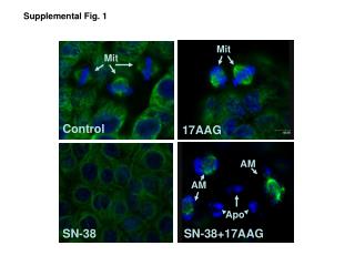
Mit Mit Control 17AAG AM AM Apo SN-38 SN-38+17AAG Supplemental Fig. 1
Supplemental Fig. 2 Tubulin
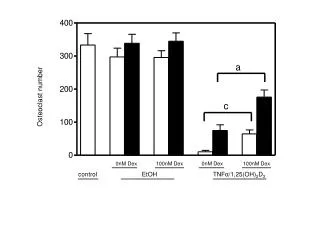
Supplemental Fig. 3 17-AAG, nM 0 100 200 500 1000 Chk1 Wee1 Tubulin MKN-74 cells
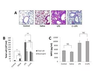
Supplemental Fig. 4 SNND ND SN24h SN17AAG 3h 6h 9h 12h 24h 3h 6h 9h 12h 24h chk1 myt1 tubulin