Download
1 / 18
200 likes | 436 Vues
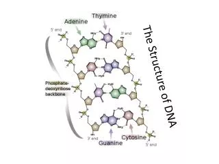
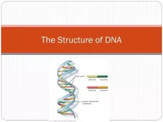
Structure of the 4 bases found in DNA. Base + sugar is called a nucleoside Base + sugar + phosphate is called a nucleotide. Structure of A-T and G-C base pairs. Hydrogen-bonds are shown as dotted red lines. A-T base pairs have 2 and G-C base pairs have 3 H-bonds. H-bonds

E N D
Structure of the 4 bases found in DNA Base + sugar is called a nucleoside Base + sugar + phosphate is called a nucleotide
Structure of A-T and G-C base pairs Hydrogen-bonds are shown as dotted red lines. A-T base pairs have 2 and G-C base pairs have 3 H-bonds H-bonds are shown as thin flat white disks in the center
3’ 5’ 5’ 3’ DNA strands are anti-parallel The first proof was provided In 1961 by measuring the ratio of different dinucleotides in DNA. The concentration of 5’AG3’ was equal to 5’CT3’ (as expected from an antiparallel orientation) and not equal to 5’TC3’ (as expected from a a parallel orientation). DNA sequencing in 1970s confirmed this conclusion.
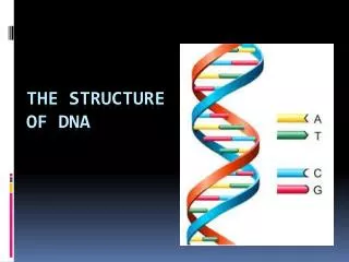
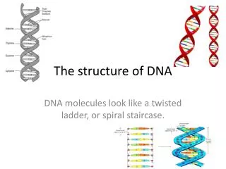
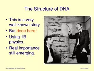
Structure of DNA Watson and Crick in 1953 proposed that DNA is a double helix in which the 4 bases are base paired, Adenine (A) with Thymine (T) and Guanine (G) with Cytosine (C).
전기영동 - +
Size separation of DNA fragments by electrophoresis in agarose gels DNA is negatively charged due to phosphates on its surface. As a result, it moves towards the positive pole.
전기영동 * Agarose gel 전기영동 시 DNA의 이동에 영향을 주는 요인들 1) DNA 분자의 크기 느리게 이동한다. 2) Agarose의 농도 느리게 이동한다. 3) DNA 형태(구조)에 따라 이동속도가 다르다. supercoiled DNA, linear DNA, open circular DNA의 순으로 빠르게 이동한다. 4) 부하되는 전압이 이동속도가 빠르다. 5) 전기장의 방향도 이동속도에 영향을 미친다. 6) Ethidium bromide는 DNA 이동속도를 15% 정도 감소시킨다. 7) 전기영동 완충용액의 성분과 이온강도도 전기영동 속도에 영향을 준다.
Distance migrated by a DNA fragment in a gel is related to log10 of its size
Plasmid DNA analysis pCDNA3.1 vector
PCR analysis H4K20me3 H3K9me3 SUV420 DNMT1 KAP-1 Input H3Ac IgG # 386 MCF7 MB231 # 411 MCF7 MB231
Genomic DNA analysis 2 uL 4 uL 8 uL Size Marker • + - + - + • + + + + + + Sonication RIPA lysis
전기영동 2) DNA 전기영동