Download
1 / 27
270 likes | 477 Vues
Overview. Spatial arrangement is called its conformation.Any structural state that is achieved without breaking covalent bonds.Most likely conformation one with the lowest ?G (native confirmation). Protein Stability. Stability tendency to maintain a native confirmation* energy difference bet

E N D
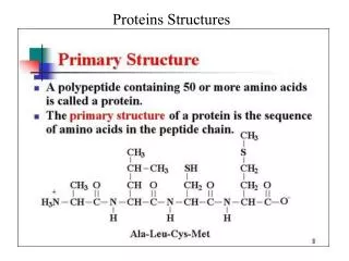
1. Chapter 6: Three-Dimensional Structure of Proteins 3-D Structure determined by AA sequence
Function = Structure
Isolated protein has unique or nearly unique structure
Most important stabilizing forces are noncovalent
Common structural patterns
2. Overview Spatial arrangement is called its conformation.
Any structural state that is achieved without breaking covalent bonds.
Most likely conformation � one with the lowest ?G (native confirmation)
3. Protein Stability Stability � tendency to maintain a native confirmation
* energy difference between folded and unfolded is 20 � 65 kJ/mol.
* entropy and hydrogen bonding tend to maintain unfolded state.
Bond strength: covalent ? 200 � 460 kJ/mol
noncovelant ? 4 � 30 kJ/mol
4. Peptide Bond is Planar and Rigid Six atoms of the peptide group lie in a single plane.
5. Secondary Structure Local conformation of some part of the polypeptide.
a. a-helix
b. �-sheet
Predicted in 1951 by Pauling and Corey 7 years before first structure elucidated.
6. Secondary Structure (Cont�d) Hydrogen bonds involving carbonyls and amines.
e.g. a-keratin � regular structure repeats every 5.15 to 5.2 Angstroms.
Helix structure is simplest structure rigid backbone can achieve.
* each turn roughly 3.6 AAs residues.
* turns always right-handed.
* about � of all AA residues in PPs are found in helices.
7. Helices (Cont�d) Optimal use of H-bonds
With helix, every peptide bond involved in H-bond
Form with either D or L AAs
* must all be same stereoisomer
8. Stability of Helices Affected by AA sequence
Long block of Glu residues will not form H-bonds (repulsive forces); same with Arg or Lys
Bulk and shape of Asn, Ser, Thr, and Cys tends to inhibit helix formation
Proline � rigidity of peptide linkage, lack of hydrogen on N
Gly: conformational flexibility; polymers of Gly tend to form coiled structures different than a-helices.
9. 5 Factors Affecting Stability of Helices Electrostatic attraction or repulsion between successive AAs
Bulkiness of adjacent R groups
Interactions between residues 3 � 4 positions apart
Occurrence of Pro and Gly
Interaction between residues at ends of helix
10. ?-Sheets Backbone is zig-zag structure instead of helix
Arranged side by side to form a series of pleats held together by H-bonds
Adjacent sheets can have parallel or anti-parallel orientation
R groups tend to be small (Gly, Ala)
11. �-Turns In globular proteins, 1/3 of AAs are in turns or loops where chain reverses direction (connect runs of helices or sheets)
?-Turns: 180o turn involving 4 AAs
* H-bond between carbonyl of 1st and amino of 4th
Gly and Pro � flexibility; imino N of Pro readily adopts cis confirmation
Found near surface, 2 inner residues form H-bond with water.
Table 6-10
12. Tertiary and Quaternary Structures Tertiary � overall 3-D structure (folding)
*Bend-producing residues: Pro, Thr, Ser, and Gly
Quaternary � arrangement of protein subunits
1. Fibrous proteins � arranged in strands or sheets
2. Globular proteins � spherical or globular shape.
13. Fibrous and Globular Fibrous: largely single type of secondary structure
Globular: several types of secondary structures
Fibrous: support, shape, and external protections
Globular: enzymes and regulatory functions
14. a-Keratin, Collagen and Silk Give strength and flexibility ? single repeating element of secondary structure
Fibrous proteins are insoluble in water (hydrophobic residues)
15. a-Keratin Hair, wool, nails, claws, quills, horns, hooves and much of the outer layer of skin
Intermediate filament (IF) proteins
Right-handed helix
* Watson and Pauling predicted coiled coil.
* Two strands oriented parallel
*Helical path of supertwist is left-handed
16. a-Keratin (cont�d) Surfaces that are in contact made up of hydrophobic residues (R groups meshed together in a regular interlocking pattern)
* Close packing of chains
* Ala, Val, Leu, Ile, Met, and Phe
Tertiary Structure: dominated by alpha helical secondary structure with its helical axis twisted in a left-handed superhelix.
Refer to picture on page 171
17. Collagen Connective tissue such as tendons, cartilage, the organic matrix of bone, and the cornea
Helix is left-handed and 3 AA residues per turn
Coiled coil: 3 separate PPs (alpha chains) supertwisted around each other to form a right-handed superhelix
18. Collagen (cont�d) 35% Gly, 11% Ala, and 21% Pro and HyPro
* Gelatin: low in essential AAs
* Repeating tripeptide unit: Gly-X-Pro or Gly-X-HyPro where X can be any residue
* Only Gly fits in tight junctions between chains
* Close packing of PP chains
See picture at bottom of page 173.
19. Collagen (cont�d) Chains cross-linked by unusual types of covalent bonds (Lys, HyLys, or His)
See picture on page 174.
Typical mammal has more than 30 variants of collagen that occur in different tissues.
Osteogenesis imperfecta � abnormal bone formation in babies
Ehlers-Danlos Syndrome � loose joints
* Cys or Ser for Gly ? disrupt repeat unit
20. Silk Fibroin Produced by insects and spiders in PP chains predominantly in the beta conformation
Rich in Ala and Gly residues (close packing of beta sheets and interlocking arrangement of R groups).
See picture on page 174.
Stabilized by extensive H-bonding between all peptide linkages
* does not stretch � beta sheets fully extended.
* held together by weak interactions - flexible
21. Globular Proteins Enzymes, transport proteins, motor proteins, regulatory proteins, etc
Protein Data Bank (PDB)
22. Myoglobulin John Kendrew et al. (1950s)
Oxygen binding protein in muscle cells
* stores oxygen and facilitates oxygen diffusion in rapid muscle movement
Single PP � 153 Aas and single iron protoporphyrin (heme)
Discuss pictures on page 176
23. Myoglobin (cont�d) Abundant in diving mammals (brown muscles)
Longest helix (23 AAs), shortest (7 AAs)
>70% of AAs in helices
Hydrophobic R groups in interior
Hydrophilic (polar) on the surface and hydrated
24. Myoglobin (cont�d) All peptide linkages in planar trans conformation
3 of 4 Pro residues are at bends
4th is in helix ? creates kink for packing
Ser, Thr, Asn ? in bends
Fe has two binding sites: one to His (93) and one to oxygen
25. Common Structural Patterns in Globular proteins Supersecondary structures: motifs or folds
* particularly stable arrangements of several elements of secondary structure.
26. Loss of Protein Structure Results in Loss of Function Loss of 3-D structure sufficient to cause loss of function is called denaturation
See Figure 6-26
Not necessarily complete unfolding
Heat (loss of H-bonds) � onset is rapid
Extremes of pH, organic solvents, detergents
No covalent bonds are broken
Can be reversible (ribonuclease)
27. Assisted Folding Not all PP fold spontaneously ? molecular chaperones assist in folding
* Hsp70 � cells stressed by high temps.
* chaperoins � protein complexes (necessary for growth of certain bacterial viruses)
Protein disulfide isomerase (interchange or shifting of disulfide bonds)
Peptide prolyl cis-trans isomerase (catalyzes isomerization of Pro)