Evolution and Optimization of Respiratory Systems in Vertebrates
Explore the coevolution of circulatory and respiratory systems in vertebrates, leading to the development of efficient gas exchange mechanisms. Learn about respiratory organs and the process of respiration at a systemic and cellular level. Discover how evolution has optimized gas exchange and the mechanics of breathing in terrestrial animals.


Evolution and Optimization of Respiratory Systems in Vertebrates
E N D
Presentation Transcript
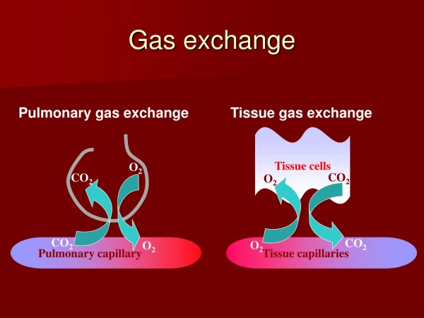
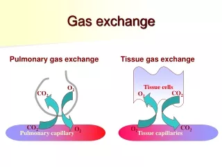
Gas Exchange By Zoe Kopp-Weber
Coevolution of circulatory and respiratory systems • Allowed for vertebrates to develop larger bodies and locomotion. • As these abilities grew, the need for efficient delivery of nutrients and O2 and removal of wastes and CO2 from the growing mass of tissues grew too.
Coevolution of circulatory and respiratory systems (cont.) • Gills developed in fish and with it the 4-chamber heart, one of the major evolutionary innovations in vertebrates. • Mammals, birds and crocodiles also have a 4-chamber heart, with 2 separate atria and 2 separate ventricles. • Right atrium receives deoxygenated blood and sends it to right ventricle which pumps blood to lungs. Left atrium receives oxygenated blood and delivers it to the left ventricle to pump the blood to the rest of the body.
For most multicellular animals, gas exchange requires special respiratory organs which provide intimate contact between gases in the external environment and the circulatory system.
Respiration describes the uptake of O2 from the environment and disposal of CO2 into the environment at a body system level. • Cellular respiration = internal respiration • Gas exchange = external respiration • Communication between internal and external respiration is provided by the circulatory system.
Respiration involves processes ranging from the mechanics of breathing to the exchange of O2 and CO2 in respiratory organs. • Respiratory organs • Invertebrates: epithelium, trachae and gills • Fish and larval amphibians: gills • Other amphibians: skin or epithelia used as supplemental/primary external respiratory organ. • Mammals, birds, reptiles, adult amphibians: lungs
Respiration involves the diffusion of gases across the plasma membrane • Which must be surrounded by water to be stable. • Thus the external environment is always aqueous, even in terrestrial animals.
Rate of diffusion between 2 sides of the membrane has a relationship called Frick’s Law of Diffusion • R=D x A delta p/d • R= rate • D= diffusion constant • A= area diffusion occurs • Delta p= difference in concentration btw interior of organism and external environment • d= distance across diffusion occurs
Evolution has optimized R via increased surface area, decreased distance and increased concentration difference. • Levels of O2 required can’t be obtained by diffusion alone over distances greater than 0.5 mm. • Vertebrates decreased this distance through the development of respiratory organs and bringing the external environment closer to the internal fluid
Dry air is composed of 78.09% N, 20.95% O2, 0.93% Ar and other inert gases, and 0.03% CO2. • This composition remains constant at altitudes of at least 100 km but the amount of air decreases as the altitude goes up. • Humans don’t survive long over 6000 meters, though the same composition of O2 is there, the atmospheric pressure brings it to only half the amount of 02 than what’s at sea level.
Though gills are effective in aquatic environments, there are two reasons terrestrial animals replaced gills with other respiratory organs. • 1. Air is less buoyant than water. Gills collapse out of water while internal air passages remain open because the body provides structural support. • 2. Water diffuses into air via evaporation. Terrestrial animals are constantly surrounded by air and therefore lose H2O. Gills would provide a large surface area for H2O loss.
Terrestrial respiratory organs • Trachae – used by insects and is a network of air-filled tubular passages. • Lung – moves air through branched tubular passages. Air is saturated with H2O before reaching a thin, wet membrane that allows gas exchange. • All but birds use a uniform pool of air • Moves in and out of the same airway passages
Mammals have higher metabolic rates so they require a more efficient respiratory system. • Lungs are packed with tiny, grape-like sacs called alveoli. Air is inhaled through mouth/nose, past the pharynx to the larynx where it then passes through the glottis and into the trachea.
The trachea splits into right and left bronchi which enter into each lung and subdivide into bronchioles that deliver air into the alveoli. • All gas exchange btw air and blood occurs across walls of alveoli.
Visceral pleural membrane – a thin membrane that covers the outside of each lung. • Parietal pleural membrane – lines the inner wall of the thoracic cavity. • Pleural cavity – the space between these two membranes, very small and filled with fluid. • Fluid allows membranes to adhere to each other, coupling the lungs to the thoracic cavity. • Pleural membranes package each lung separately so if one should collapse, the other can function.
Mechanics of breathing • In all terrestrial vertebrates but amphibians, air is drawn into the lungs by subatmospheric pressure. • Boyle’s Law – when the volume of a given quantity of gas increases, its pressure decreases. • When inhaling, volume of thorax is increased and the lungs expand. Lowered pressure in lungs allows air to enter.
Diaphragm – a muscle that increases thoracic volume by contracting. • When it contracts, it assumes a flattened shape and lowers, expanding the volume of the thorax and lungs while adding pressure onto the abdomen. • External intercostal muscles – also contributes in increasing thoracic volume. • These muscles between the ribs contract, causing the ribcage to expand.
The thorax and lungs have a degree of elasticity. • They resists distension and recoil when distending force subsides.
Breathing measurements • At rest, each breath moves a tidal volume of 500 mL of air in and out of the lungs. • 150 mL in trachea, bronchi and bronchioles where no gas exchange occurs. • Anatomical dead space, air here mixes with fresh air during inhalation. • Maximum amount of air expired after a maximum inhalation is called the vital capacity. • Averages 4.6 liters in young men and 3.1 liters in young women.
Hypoventilating - when breathing is insufficient to maintain normal blood gas measurements. • Hyperventilating – when breathing is excessive for a particular metabolic rate. • Increased breathing after exercise isn’t necessarily hyperventilating because faster breathing is matched to faster metabolic rate and blood gas measurements remain normal.
Mechanism regulating breathing • Each breath initiated by a respiratory controntrol center in the medulla oblongata. • Neurons send impulses that stimulate muscles to contract and expand the chest cavity. • Though controlled automatically, these controls can be overridden by, for example, holding one’s breath.
A fall in blood pH stimulates neurons in aortic and carotid bodies • These are sensory structures known as peripheral chemoreceptors in the aorta and carotid artery. • Send impulses to the respiratory control center in the medulla oblongata, which stimulates increased breathing. • responsible for immediate stimulation when the blood partial CO2 pressure rises.
Central chemoreceptors – responsible for sustained increase in ventilation if partial CO2 pressure remains elevated. Increased respiratory rate acts to eliminate extra CO2, bringing blood pH to normal.
Hemoglobin and gas transport • When O2 diffuses from alveoli into blood, the circulatory system then delivers the O2 to tissues for respiration and carries away the CO2. • Amount of O2 dissolved in blood plasma depends directly on the partial O2 pressure or the air in the alveoli. • When lungs function normally, the blood plasma leaving the lungs have almost as much DO as possible. • Whole body carries almost 200 mL/L of O2, most is bound to molecules of hemoglobin inside red blood cells.
Hemoglobin - protein composed of four polypeptide chains and four organic compounds (heme groups). • Each heme group has an iron atom at the center, able to bind to a molecule of O2. • Allows hemoglobin to carry four molecules of O2.
Hemoglobin loaded with O2 forms oxyhemoglobin. • Bright red, tomato juice color • As blood passes capillaries, some oxyhemoglobin releases oxygen, becoming deoxyhemoglobin • Dark red but gives tissues a bluish tinge.
Red color, oxygenated • Blue color, oxygen-depleted
Hemoglobin is used by all vertebrates, and also by many invertebrates • Other invertebrates use hemocyanin as an oxygen-carrier • O2 binds to copper rather than iron. • Not found in blood cells but rather dissolved in circulating fluid of invertebrates
Oxygen transport • As blood travels through the systemic blood capillaries, O2 leaves the blood and diffuses into tissues. • 1/5 of O2 is unloaded in tissues, 4/5 in blood as a reserve. • The reserve allows the blood to supply the body O2 during exercise. • Also ensures enough O2 to maintain life 4-5 minutes if breathing is interrupted or the heart stops.
O2 transport affected by • CO2: produced by metabolizing tissues, it combines with H2O forming carbonic acid. This dissociates into bicarbonate and H+, lowering blood pH. • Also reduces hemoglobins affinity for O2 and causes it to release O2 more readily. • This is all called the Bohr effect. • Increase in temperature has a similar effect. • Skeletal muscles produce CO2 quicker during exercise, producing heat.
Carbon Dioxide Transport • Systemic capillaries deliver O2 and remove CO2 from tissues • Majority diffuses into red blood cells where it’s catalyzed with water to form carbonic acid (H2CO3) • Disassociates into bicarbonate and H+ and moves into the plasma, exchanging a chloride ion for a bicarbonate (chloride shift). • Removes large amounts of CO2 from plasma, facilitating diffusion of additional CO2 into plasma from surrounding tissues.
Blood carries CO2 to the lungs in this form. • CO2 diffuses out of red blood cells, into the alveoli and then leaves the body with exhalation.
Nitric Oxide Transport • Nitric oxide acts on many cells to change their shape/functions. • Causes blood vessels to expand by relaxing surrounding muscle cells. • Blood flow/pressure regulated by nitric oxide in bloodstream.
One hypothesis proposes hemoglobin carries super nitric oxide which is able to bind to cysteine in hemoglobin • Dumps CO2 and picks up O2 and NO in the lungs • To increase blood flow, hemoglobin can release super NO into blood, making blood vessels expand • Can also trap excess NO on vacant iron atoms, making blood vessels constrict. • Red blood cells return to lungs, hemoglobin dumps CO2 and regular NO. Then ready to picl up O2 and super NO.
Disease • Emphysema - usually caused by cigarette smoking, the vital capacity of the lungs is reduced and alveoli are destroyed. • Bronchitis - a respiratory infection affecting nose, sinus and throat, then moves on into the lungs. Cough produces an excess of mucus. • Pneumonia - inflammation of the lungs that can be caused by bacteria, viruses or fungi. Causes coughing, fever and it will likely make it harder to breathe.