Nervous System (NS) – Cerebral Cortex
بسم الله الرحمن الرحيم. Dr. Othman Al- Shboul Department of Physiology. Nervous System (NS) – Cerebral Cortex. Based on differences in the structure, location, and functions, nervous system is subdivided into: Central nervous system (CNS) Brain and spinal cord Contained within bone

Nervous System (NS) – Cerebral Cortex
E N D
Presentation Transcript
بسم الله الرحمن الرحيم Dr. Othman Al-Shboul Department of Physiology Nervous System (NS) – Cerebral Cortex
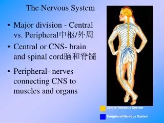
Based on differences in the structure, location, and functions, nervous system is subdivided into: • Central nervous system (CNS) • Brain and spinal cord • Contained within bone • Peripheral nervous system (PNS) • All nerve tissue outside CNS • Sensory & motor divisions Organization of the Nervous System
Generalized Model of Function of NS INPUT (PNS: sensoryneurons; afferentneurons) INTEGRATION (CNS: inter-neurons) OUTPUT (PNS: motor neurons; efferent neurons)
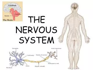
Cells of the Nervous System • Consists of 2 kinds of cells: • Neurons: functional units of NS • Supporting cells (= glial cells) • Maintain homeostasis • Are 5X more common than neurons • Schwann and satellite cells in the PNS • Oligodendrocytes, microglia, astrocytes and ependymal cells in the CNS
Afferent neurons: • Sensory receptor at peripheral ending (generates AP) • Convey input to the CNS • Cell body outside CNS • Efferent neurons: • Cell bodies in the CNS • Convey output for the effector organs (muscles, glands) • Interneurons: • 99% of all neurons • Integrating peripheral responses to peripheral information Functional Classes of Neurons
Consists of: • The brain • Spinal cord CNS
Major brain functions: • Regulates internal environment • Experiences emotions • Voluntarily controls movements • Perceives own body and surroundings • Engages in other higher cognitive processes (e.g. thought and memory) • Brain functions as a whole (neurons linked via synapsis). The Brain
Based on anatomical distinction, functional specialization, and development, brain has the following regions: • Brain stem (Medulla, pons, midbrain) • Cerebellum • Forebrain • Diencephalon • Hypothalamus • Thalamus • Cerebrum • Basal nuclei • Cerebral cortex Major Components of the Brain
the outermost sheet of neural tissue (gray matter) of the cerebrum of the brain • Total surface area: 2200 cm2 (2.5 ft2) • Thickness: 1.5 mm - 4.5 mm • Weight: 600 gm (40 % of total brain weight) • 180 gm --------- neurons (10-15 billion neurons) • 420 gm --------- glial cells • Function: motor control of the body & information processing center Cerebral Cortex neuronal cell bodies &their dendrites & most glial cells Myelinated nerve fibers (axons)
Functions of Cortical Lobes • Voluntary motor activity • Speaking ability Receiving and processing sensory input Initial processing of visual input sound (auditory) sensation
Somatosensory Cortex • The somatosensory cortex: • located in the front portion of each parietal lobe • Immediately behind the central sulcus • Post-central gyrus • It is the site for initial cortical processing • and perception of: • Somestheticinput (sensations from the surface of the body, such as touch, pressure, heat, cold, and pain ) • Proprioceptive input (awareness of body position)
Somatosensory Cortex • Each region within the somatosensory cortex receives input from a specific area of the body. • Different parts of the body are not equally represented • The size of each body part in this homunculus indicates the relative proportion of the somatosensory cortex devoted to that area. (e.g., fingers >>> trunk) • Reception of opposite side inputs Sensory Homunculus Proportional representation of the different body parts and areas in brain hemispheres
Somatosensory Cortex • Thalamus vs. somatosensory cortex: • Thalamus simple awareness of sensation • Somatosensory cortex full sensory perception (location, intensity ….) • Capable of spatial discrimination; shapes and small differences between objects • Connections with higher brain centers: • via white matter fibers to adjacent higher sensory areas for furtherelaboration, analysis, and integration of sensory information.
Primary Motor Cortex • Primary motor cortex: • Immediately in front of the central sulcus • Pre-central gyrus • Next to the somatosensory cortex • Confers voluntary control over movement produced by skeletal muscles. • Controls opposite side muscles of the body
Primary Motor Cortex • The extent of representation in the motor cortex is proportional to the precision and complexity of motor skills required of the respective part • E.g., lips are >>> trunk area Motor Homunculus Proportional representation of the different body parts and areas in brain hemispheres
When an area of the brain associated with a particular activity is destroyed, other areas of the brain may gradually assume some or all of the functions of the damaged region • Mechanism: ???formation of new neural pathways (not new neurons, but new connections between existing neurons) Brain Plasticity
The recording of electrical activity (potentials) along the scalp • A tracing (measurement) of voltage fluctuations resulting from ionic current flows within the neurons of the brain versus time recorded from electrodes placed over scalp in a specific array • Deep parts of the brain are not well sampled Electroencephalogram (EEG)
EEG Elements • Electrodes: • Active electrodes: Attached to the scalp • Reference electrode: Mastoid, nose, ear lobe... • Amplifier The EEG records differences in voltage – difference in electrical potential from one electrode to another
EEG Rhythms • Alpha (α) waves • Most common in adults. • Posteriorly (occipital) more than anteriorly • Especially prominent with closed eyes and with relaxation. • Disappears normally with attention (e.g., mental arithmetic, stress, opening eyes). • In most instances, it is regarded as a normal waveform.
EEG Rhythms • Beta (β) waves • Small in amplitude • More evident anteriorly • Drugs, such as barbiturates and benzodiazepines, augment beta waves
EEG Rhythms • Theta (θ) waves • Normally seen in sleep • In awake adults, these waves are abnormal if they occur in excess.
EEG Rhythms • Delta (δ) waves • Normally seen in deep sleep. • Delta waves are abnormal in the awake adult. • Often, they have the largest amplitude of all waves. • Theta and delta waves are known collectively as slow waves.
To distinguish various stages of sleep A clinical tool in the diagnosis of cerebral dysfunction (e.g. Epilepsy) Legal determination of brain death EEG Uses