PELVIC FRACTURES
620 likes | 1.1k Vues
PELVIC FRACTURES. An overview Dr.Abraham John, Specialist,A/E, Al-Khor Hospital. The problem. Devastating Injuries with many complications and require extensive rehabilitation. About 0.3-6% of all fractures and seen in upto 20% of polytrauma cases. Incidence.

PELVIC FRACTURES
E N D
Presentation Transcript
PELVIC FRACTURES An overview Dr.Abraham John, Specialist,A/E, Al-Khor Hospital
The problem • Devastating Injuries with many complications and require extensive rehabilitation. • About 0.3-6% of all fractures and seen in upto 20% of polytrauma cases.
Incidence • Bimodal Age Distribution: 15-30 years(High Energy) and 50- 70 years(Low Energy) • Average age 31.5 years • Male predominance (57-75%) • Mortality rates vary from 6-40% (depending on the type of # and associated injuries)
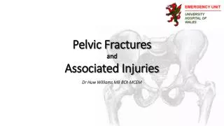
Anatomy of the Pelvis: Outline • Joins the axial and appendicular skeletons • Transfers weight from the vertebral column to the lower limbs when standing
Anatomy: Bony • 3 Bones: Ilium, Ischium, Pubis –joined at the acetabulum (triradiate cartilage) • Sacrum(5 fused Vx) • Coccyx(4 fused Vx) • BONES OF PELVIS HAVE NO INHERENT STABILITY
Anatomy- Ligamentous • Ligaments provide stability to the Pelvis • Strong ligaments –div.into 4 groups 1.Anterior and 2.Posterior S.I. Ligaments 3.Sacrotuberous and sacrospinous 4. Ligs. of pubic symphysis
Vascular anatomy of the pelvis 1.Iliolumbar A 2.Lat.Sacral Art. 3.Sup Gluteal Art. 4.Obturator Art. 5.Int.Pudendal Art. 6.Inf Gluteal Art. 7.Vesical Art
Pathophysiology of Pelvic # • Pelvic # results from 3 vectors primarily: 1.AP Compression (55-70%) eg. Head on MVC -passenger 2.Lateral compression (13-15%) eg.Side impact MVC, Pedestrian 3.Vertical shear (7-15%) eg.Fall from height
Mech. Of Injury: AP compression AP Fracture Patterns: A: ST and SS Ligaments intact, B:Only Post.SI ligament intact C:Complete disruption AP Mechanism
Mech. Of injury: Lat compression Fracture Patterns in Lat. Compression Injury
Classification of Pelvic Fractures Pelvic Fractures are classified acc. to the direction of the injuring force. 2 major systems used : 1. Tile system- Focuses on pelvic stability 2. Young and Burgess system – Focuses on the degree of injury
Tile Classification of Pelvic # Type A : Posterior Arch Intact - #s of Pelvic Ring-pubic rami or iliac wing, Transverse sacral # • Stable Fractures • Subdivided into A 1,2,3 and each into 3 • No hemipelvis displacement , insignificant displacement of individual bony fragments
Tile Classification of Pelvic # • Type A : Posterior Arch Intact A2-min.displacement A3-Transverse sacral A1-avulsion injury
Tile Classification of Pelvic # • Type B :Incomplete Post.Arch Disruption -Cause : AP or Lateral compression or combined force -External or Internal rotational deformity of the hemipelvis -Rotationally unstable, vertically stable
Tile Classification of Pelvic # • Type B :Incomplete Post.Arch Disruption B2- Lat.comprsn, int.rotated B1-AP comprsn, unilat B3-Bilat rotational instability
Tile Classification of Pelvic # • Type C: Complete Post. Arch Disruption -Vertical Shear with or without assoc. AP/Lat compression injury -Ipsilateral or Bilateral hemipelvis vertical (cranial) displacement. -Vertical and Rotational Instability
Tile Classification of Pelvic # • Type C: Complete Post. Arch Disruption C2-Bilateral: one side Vertical and other side Rotational instability C3- Bilateral rotational instability C1-unilateral Xray/ CT image of Symphyseal disruption- Lt S.I. Jt disruption
Evaluation • Should be meticulous, along with resuscitation according to ATLS guidelines. • Assess pelvic stability gently once by AP and Lat. Compression.(May induce fresh bleeding) • Assess leg rotation and length ,look for pelvic/flank ecchymosis, scrotal/vulvar swelling • Look for Blood at the urethral meatus, High riding prostate,distended bladder- s/o Urethral Injury. • Rectal and Vaginal Exam mandatory.
Investigations RADIOGRAPHY : --AP radiograph of the Pelvis is an adjunct to the Primary Survey --Oblique (Judet ) views are done in suspected Acetabular Fractures --Inlet and Outlet views are done in Pelvic Ring Fractures ( Inlet View- Xray Tube At patients Head end and angled 45deg. Beam perpendicular to the brim Outlet View –Tube at the feet end and angled up 45 deg –beam perpendicular to the sacrum) AP View Inlet View Outlet View
Investigations- Pelvic CT • Good Sensitivity and Specificity • Subtle # and disruptions seen better • Axial/Sagittal/Coronal plane images can be seen. Reformatting into 3D images to view acetabular injuries possible • Pelvic soft tissue can be examined. • Limitation:Patients need to be transported to the CT room
C T Images of pelvic # Sacral Buckle Fracture (Lat Comp) AP comp Injury- Ext Rotation & Lt Si Jt Verical shear: SI jt disrupted ant & posteriorly 3D CT showing Ant and Post disruption
Other Investigations: • Pelvic Angiography: Diagnostic to localise Vascular injury and Therapeutic for embolisation MRI, Ultrasound and Radionuclide studies have no role in the evaluation of Pelvic Fractures alone.(FAST is exception)
G.U.Injury Assessment • Injuries to Bladder and Urethra most common • Blood at the meatus, inability to void , high riding prostate on PR S/O urethral injury. • Retrograde Urethrogram and Cystogram indicated in suspected injury
Management of Pelvic Fractures • According to the ATLS guidelines • ABCDE evaluation- • Hemodynamically unstable patient with an unstable pelvis – Resuscitation with Crystalloid infusion with R/L thru 2 wide bore cannulae, Blood Transfusion, AP x-ray of the pelvis, DPL if required, Pelvic stabilisation with sheets, PASG CT evaluation,Angiography for diag. And Embolisation Urgent Ortho.consultation for Ext.Fixation
Fixation Techniques • These help to stabilize the pelvis, reduce pelvic volume and control bleeding. 1.External Fixation 2.Pelvic Clamp 3.Traction 4.Sling application with int.rotation of the extremities.
Pelvic Angiography Used to treat potentially life threatening Haemorrhage from Pelvic fractures. This demonstrates the vessels responsible for the bleeding and facilitates embolisation Ant..Division of Int Iliac Cut off Coils In Place
Associated injuries Pelvic Fractures are rarely found in Isolation. The associated Injuries found commonly are: *Closed Head injury--------51% *Long Bone Fracture-------48% *Peripheral Nerve Injury –26% *Thoracic Injury-------------20% *Urethral Injury--------------18% *Bladder-----------------------10% *spleen----------------------- 10% *Liver---------------------------7% *Other (GIT, Mesentery,Kidney,Fe Urethra)—21%
Treatment • Depends on Fracture type and associated injuries • Multi disciplinary approach involving Trauma surgeon,Orthopaedic surgeon, Interventional radiologist for embolisation • High Energy Fractures (Type B and C) initially treated with Ext.fixation and then if required ORIF for unstable injuries. • Early mobilisation aimed for and full weight bearing expected in 3 months • Low energy fractures (Type A) treated symptomatically with pain relief and weight bearing as tolerated. Surveillance X-rays are taken 11-2 weeks after injury and initiating weight bearing to confirm maintenance of stability.
Complications Immediate: 1)Pelvic Bleeding: A)Venous-from post. pelvic veins, marrow of bones. B)Arterial-Due to direct arterial injury Any branch of the int. iliac depending on site of fracture. 2)Bladder Injury- Extra or intraperitoneal 3)Urethral injury- Membranous urethra injured most as it passes thru the urogenital diaph. 4)Nerve Injury-fractures of the sacrum may damage the sacral plexus, Sciatic Nerve Injury in fractures involving the acetabulum
Complications- Early • Continued Blood Loss leading to shock, renal failure,coagulopathy etc • Infection –Pelvic haematoma may get infected leading to abscess formation • Thromboembolic phenomena may occur at any time with venous thrombosis in the lower limbs or the pelvic veins. Doppler ultrasonography used to evaluate the venous system. Pelvic veins are seen better with magnetic resonance venography.
Complications-Late • Pain is the most common long term complaint of patients • Malunion of fractures causing limb length discrepancies, gait abnormalities and pain. • Non union of fractures – usually seen in vertically unstable fractures
Prevention • High Energy Fractures: Education,Safety and Law enforcement are the most important factors in prevention. • Low Energy Fractures: Prevention and treatment of Osteoporosis is the most effective .