Download
1 / 56
580 likes | 812 Vues
Lecture VIII. The Spinal Cord, Reflexes and Brain Pathways. Bio 3411 Monday September 27, 2010. Readings. NEUROSCIENCE 4 th ed: Review Chapter 1 pp. 11-22; Read Chapter 9 pp. 207-212, 218 Study Box 9A, Figure 9.8 & Refer to Table 9.1; Read Chapter 16 pp. 399–414

E N D
Lecture VIII. The Spinal Cord, Reflexes and Brain Pathways Bio 3411 Monday September 27, 2010
Readings NEUROSCIENCE 4th ed:Review Chapter 1 pp. 11-22; Read Chapter 9 pp. 207-212, 218 Study Box 9A, Figure 9.8 & Refer to Table 9.1; Read Chapter 16 pp. 399–414 Study figures 16.2,16.3,16.4, 16.14 Read Chapter 17 pp. 432–436 Study figure 17.9 THE BRAIN ATLAS 3rd ed: Read pp. 4-17 on class web site Look at pp 36, 43, 49, 75-76, 140, 151, 154, 170-171, 182-183, 200-201. Lecture VIII. The Spinal Cord, Relexes and Brain Pathways
What the last Lecture was about • The Initiation of the Central Nervous System • CNS Growth and Pattern Development • Bug Brains • Several Mechanisms for Directing the Show (scripts conserved) • How did vertebrate and invertebrate patterns arise? • Reprise and overview the discussions of developmental sequences and mechanisms in the nervous system Lecture VIII. The Spinal Cord, Relexes and Brain Pathways
Overview for this Lecture Spinal Cord Columns, Horns, Spinal Segments Spinal Nerves Dermatomes, Motor Units Reflexes “Knee Jerk”- Myotatic or Stretch Reflex Withdrawal & Crossed Extensor Reflexes Two Spinal Pathways Sensory - Dorsal Column/ Medial lemniscus Motor - Cortico-spinal Tract Lecture VIII. The Spinal Cord, Relexes and Brain Pathways
Cervical (C) - Neck Thoracic (T) - Chest Lumbar (L) - Back Sacral (S) - Pelvis (The Brain Atlas 3rd ed,p. 8) Segments of spinal cord, spinal nerves and vertebrae Lecture VIII. The Spinal Cord, Relexes and Brain Pathways
Human spinal cord from side, front and back ≈ 50 cm Lecture VIII. The Spinal Cord, Relexes and Brain Pathways
Internal Structure • Canal = tube • White Matter - columns, tractsanterior up and down to and from brain lateral down from brain (>>up)posterior mainly up to brain • Gray Matter - posterior (dorsal) horn “sensory”,anterior (ventral) horn “motor” Lecture VIII. The Spinal Cord, Relexes and Brain Pathways
Front (anterior/ventral) Anterior Column (white matter) Anterior Horn (gray matter) Section of human spinal cord (C8) myelin stain Lateral Column (white matter) Posterior Horn (gray matter) Back (posterior/dorsal) Posterior Column (white matter) Lecture VIII. The Spinal Cord, Relexes and Brain Pathways
Front (anterior/ventral) Anterior Horn Anterior Column Section of human spinal cord (C8) cell body stain Lateral Column Back (posterior/dorsal) Posterior Horn Posterior Column Lecture VIII. The Spinal Cord, Relexes and Brain Pathways
Cervical Cord (The Brain Atlas 3rd ed, p.151) Sacral Cord (The Brain Atlas 3rd ed, p.154) The size of white matter tracts (posterior, lateral and anterior columns) increases as more axons are added on the way TO the brain and decreases as axons end on the way FROM the the brain. Lecture VIII. The Spinal Cord, Relexes and Brain Pathways
Spinal Nerves • Intervertebral foramen • Segmental spinal nerve • Compound action potential Lecture VIII. The Spinal Cord, Relexes and Brain Pathways
The Brain Atlas 3rd ed, p. 49. Spinal canal Intervertebral foramen Left - Vertebral bones. Right - Human spinal cord in cross–section showing anterior (ventral) and posterior (dorsal) spinal roots and spinal or posterior (dorsal) root ganglion (posterior to right). Lecture VIII. The Spinal Cord, Relexes and Brain Pathways
Mixed spinal nerve Segmental nerve: (posterior (dorsal) root = sensory - touch; anterior (ventral) root = motor -movement; spinal or posterior (dorsal) root ganglion = sensory nerve cell bodies) Periphery (skin, muscle, etc.) Spinal cord front = anterior back = posterior Lecture VIII. The Spinal Cord, Relexes and Brain Pathways
George H. Bishop 1889 - 1973 Joseph Erlanger 1874 - 1965 (Prix Nobel 1944) Herbert Spencer Gasser 1888 - 1965 (Prix Nobel 1944) Lecture VIII. The Spinal Cord, Relexes and Brain Pathways
Periphery (skin, muscle, etc.) Spinal cord Mixed spinal nerve front = anterior back = posterior Stimulate (Shock) Record Lecture VIII. The Spinal Cord, Relexes and Brain Pathways
Discriminative Touch Fast pain & Temperature (cold) Slow pain & Temperature (warm) Compound action potential Erlanger, Gasser (Bishop) Conduction Velocity m/s Lecture VIII. The Spinal Cord, Relexes and Brain Pathways
Axon diameters differ in motor and sensory nerves Cross section of human muscle (motor) nerve – myelin stain Cross section of human sensory nerve – myelin stain Lecture VIII. The Spinal Cord, Relexes and Brain Pathways
Segmental Nerves Spinal orPosterior (dorsal) Root, Ganglion Cells & Sensory Nerves (axons in from posterior (dorsal) root ganglia) Dermatomes Anterior (ventral) Root & Motor Nerves (axons out from motor neurons) Motor Units Lecture VIII. The Spinal Cord, Relexes and Brain Pathways
Spinal or Posterior (dorsal) Root Ganglion Cells Pseudo-Unipolar Neurons (neurons start as bipolar cells and become “unipolar” during development) Single sensory endings light & crude touch, pain, temperature and muscle senses Axons diverge to multiple spinal targets motor neurons - c, interneurons - c, spinal cord - b, and brain -a Lecture VIII. The Spinal Cord, Relexes and Brain Pathways
Segmental nerve (posterior (dorsal) root = sensory - touch; ventral root = motor -movement; spinal or posterior (dorsal) root ganglion = sensory nerve cell bodies) Mixed spinal nerve front = ventral back = dorsal Periphery (skin, muscle, etc.) Spinal cord Lecture VIII. The Spinal Cord, Relexes and Brain Pathways
Dermatome = The region (slice) of skin innervated by a single spinal or posterior (dorsal) root ganglion Lecture VIII. The Spinal Cord, Relexes and Brain Pathways
Motor Units, Motor Neuron Pools & Somatotopy Lecture VIII. The Spinal Cord, Relexes and Brain Pathways
Spinal Motor Neurons • Multipolar • Output Diverges to - several or many muscle cells: motor unit • Input Converges from – spinal or posterior (dorsal) root ganglion cellsspinal interneuronslong tracts from from brain • Integrate • Map flexors, extensors, proximal, distal Lecture VIII. The Spinal Cord, Relexes and Brain Pathways
Section of human spinal cord (C8) myelin stain – anterior horn Section of human spinal cord (C8) cell body stain – anterior horn Lecture VIII. The Spinal Cord, Relexes and Brain Pathways
Motor Unit - A motor neuron and the muscle fibers it innervates. Lecture VIII. The Spinal Cord, Relexes and Brain Pathways
“Quads” Proximal (nearer) Extensor muscles “Shin” Distal (farther) Extensor muscles “Hamstrings” Proximal (nearer) Flexor muscles “Calf” Distal (farther) Flexor muscles Motor Neurons to Proximal (nearer) Extensor muscles Motor Neurons to Distal (farther) Extensor muscles Motor neuron pools (nuclei) are organized systematically according to the body plan - somatotopically Motor Neurons to Proximal (nearer) Flexor muscles Motor Neurons to Distal (farther) Flexor muscles Lecture VIII. The Spinal Cord, Relexes and Brain Pathways
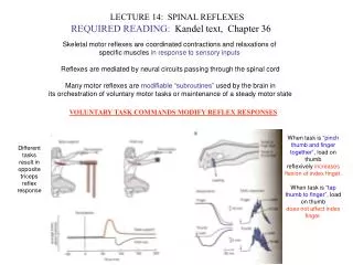
“Knee Jerk”Stretch Reflex & Antagonist Inhibition Lecture VIII. The Spinal Cord, Relexes and Brain Pathways
When the knee is struck… Ia muscle afferents fire… there is monosynaptic activation of the extensor -motor neuron… and the (agonist) muscle(s) contracts. Lecture VIII. The Spinal Cord, Relexes and Brain Pathways
When the knee is struck… Ia muscle afferents fire… there is monosynaptic activation of the extensor -motor neuron… and the (agonist) muscle(s) contracts. The knee extends. Glycinergic (inhibitory) interneurons are also activated… …which inhibit motor neurons to the flexor (antagonist) muscle. Lecture VIII. The Spinal Cord, Relexes and Brain Pathways
“Stepping on a Nail” Withdrawal & Crossed Extensor Reflexes Lecture VIII. The Spinal Cord, Relexes and Brain Pathways
Stepping on a sharp object activates pain afferents in the skin… activating interneurons in the dorsal horn… that exciteflexors and inhibit extensors… Lecture VIII. The Spinal Cord, Relexes and Brain Pathways
Stepping on a sharp object activates pain afferents in the skin… activating interneurons in the dorsal horn… that exciteflexors and inhibit extensors… and the leg flexes “withdraws.” Lecture VIII. The Spinal Cord, Relexes and Brain Pathways
But the person would fall… if the crossed extensors weren’t activated… and the crossed flexors weren’t inhibited to extend the other (contralateral) leg to stand on. Lecture VIII. The Spinal Cord, Relexes and Brain Pathways
Pathways • Subserve a particular function • Axons travel together in specific locations (i.e., tracts) in a particular order (topography) • Always consider: cell body (soma) location, axon course, synapses and side relative to origin and destination • Nomenclature often origin and target, i.e., Cortico-Spinal Tract = from cortex to spinal cord Lecture VIII. The Spinal Cord, Relexes and Brain Pathways
Path Finding • Loss of a particular function after damage (lesion) • Stimulation (natural/electrical) with recording • Pathology - degeneration of cells and axons with secondary loss of myelin • Experiments - special stains and tracers that take advantage of physiological processes Lecture VIII. The Spinal Cord, Relexes and Brain Pathways
Pathway Conventions • Related to whole brain through “sections” – gross, histological, imaging • Related to fiber bundles (fasciculi; i.e., lateral columns, internal capsule, corpus callosum) • Related to nuclei, ganglia, areas, layers • Related to transmitters and effects: excitatory, inhibitory, modulatory; fast, slower, slow Lecture VIII. The Spinal Cord, Relexes and Brain Pathways
THE BRAIN ATLAS 3nd ed, pp. 5, 7 Lecture VIII. The Spinal Cord, Relexes and Brain Pathways
Pathways - “Primitive” ––––> “Evolved’ (Synapse & Synapse Number) Intraspinal “Pathways” Midline Midline 2 1 1 Knee Jerk 1 Antagonist Inhibition 2 Lecture VIII. The Spinal Cord, Relexes and Brain Pathways
THE BRAIN ATLAS 3nd ed, p. 8 Brainstem Cerebellum Lecture VIII. The Spinal Cord, Relexes and Brain Pathways
THE BRAIN ATLAS 3nd ed, p. 151 Lecture VIII. The Spinal Cord, Relexes and Brain Pathways
THE BRAIN ATLAS 3nd ed, p. 140 Lecture VIII. The Spinal Cord, Relexes and Brain Pathways
THE BRAIN ATLAS 3nd ed, p. 75-76 Lecture VIII. The Spinal Cord, Relexes and Brain Pathways
Dorsal Column/Medial Lemniscus (a ribbon) Pathway This pathway carries fine discriminative and active touch, body and joint position, and vibration sense.
THE BRAIN ATLAS 3nd ed, p. 185 Face Hand Body Foot Lecture VIII. The Spinal Cord, Relexes and Brain Pathways
Pathways - “Primitive” ––––> “Evolved” (Synapse & Synapse Number) Intraspinal “Pathways” Ascending Sensory Pathway Midline Midline Midline 2 1 2 1 1 Knee Jerk 1 Antagonist Inhibition 2 Dosal Column/ Medial Lemniscus Pathway 2 Lecture VIII. The Spinal Cord, Relexes and Brain Pathways
Corticospinal (Pyramidal) Pathway This is the direct connection from the cerebral cortex for control of fine movements in the face and distal extremities, e.g., buttoning a jacket or playing at trumpet.
THE BRAIN ATLAS 3nd ed, pp. 36, 43 Corticospinal Tract (Pyramid) at Medulla Lecture VIII. The Spinal Cord, Relexes and Brain Pathways
Foot Hand Face All THE BRAIN ATLAS 3nd ed, p. 201 Lecture VIII. The Spinal Cord, Relexes and Brain Pathways