
NITRIC OXIDE
E N D
Presentation Transcript
NITRIC OXIDE Aseem Hussain
NITRIC OXIDE • Nitric oxide is a gas. It is highly reactive; and is one of the products in automobile exhaust and plays a major role in atmospheric pollution. • Surprisingly, it was found that it has important physiological functions.
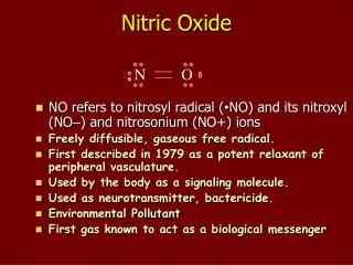
NITRIC OXIDE • This was the first discovery that a gas can act as a signal molecule in the organism. • A highly reactive compound, it only exists for six to ten seconds inside the body, then it is converted, by oxygen, into other compounds of nitrogen called nitrites. • Uniquely, it is one of the few compounds with an odd number of electrons thereby making it a ‘free-radical' prone to ionizations.
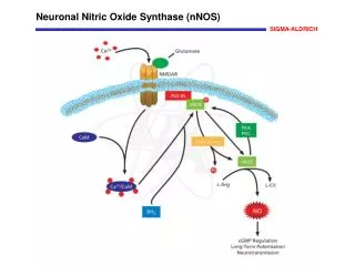
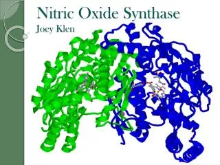
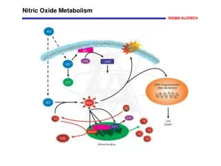
NITRIC OXIDE • NO is synthesized within cells by an enzyme NO synthase (NOS).
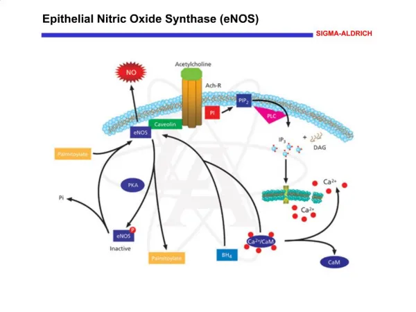
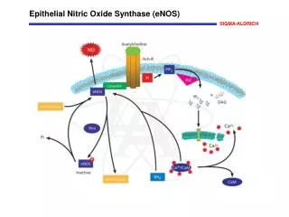
Sub-types of NOS • nNOS : found in neurons • iNOS : is inducible and found in macrophages. Whereas the levels of nNOS and eNOS are relatively steady, expression of iNOS genes awaits an appropriate stimulus (e.g., ingestion of a parasite). • eNOS : found in the endothelial cells that line the lumen of blood vessels.
NITRIC OXIDE • All types of NOS produce NO from arginine with the aid of molecular oxygen and NADPH.
NITRIC OXIDE • NO diffuses freely across cell membranes. • There are so many other molecules with which it can interact, that it is quickly consumed close to where it is synthesized. • Thus NO affects only cells near its point of synthesis.
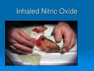
NITRIC OXIDE FUNCTIONS • Blood Flow • NO relaxes the smooth muscle in the walls of the arterioles. • Mice whose genes for the NO synthase found in endothelial cells (eNOS) has been “knocked out" suffer from hypertension. • Nitroglycerine, which is often prescribed to reduce the pain of angina, does so by generating nitric oxide, which relaxes the walls of the coronary arteries and arterioles.
Platelet aggregation • NO also inhibits the aggregation of platelets and thus keeps inappropriate clotting from interfering with blood flow
Kidney Function • Release of NO around the glomeruli of the kidneys increases blood flow through them thus increasing the rate of filtration and urine formation.
Other functions • Penile Erection • Intestinal peristalsis • Contractility of the smooth muscle wall of the uterus during labor • NO stimulates secretion from several endocrine glands. • Neurotransmitter • Memory and learning.
NITRIC OXIDE • NO aids in the killing of engulfed pathogens like bacteria within the lysosomes of macrophages • Harmless bacteria, living as commensals at the rear of our throat, convert nitrates in our food into nitrites. When these reach the stomach, the acidic gastric juice (pH ~1.4) generates NO from them. This NO kills almost all the bacteria that have been swallowed in our food.
Mechanisms of NO Action • The signaling functions of NO begin with its binding to protein receptors on or in the cell. The binding sites can be either: • a metal ion in the protein or • one of its Sulfur atoms e.g., on cysteine. • In either case, binding triggers a change in the protein which, in turn, triggers the formation of a "second messenger" within the cell. The most common protein target for NO seems to be guanylyl cyclase, the enzyme that generates the second messenger cyclic GMP (cGMP).
NITRIC OXIDE • The discovery of the biological functions of nitric oxide in the 1980s came as a complete surprise Nitric oxide was named "Molecule of the Year" in 1992 by the journal Science, • a Nitric Oxide Society was founded, and a scientific journal devoted entirely to nitric oxide was created. • The Nobel prize in Medicine in 1998 was awarded for the discovery of the signaling properties of nitric oxide.
NITRIC OXIDE • It is estimated that yearly about 3,000 scientific articles about the biological roles of nitric oxide are published. • The following article is one of these…
Nitric oxide-induced cellular stress and p53 activation in chronic inflammation • This article was published in “The Proceedings of the National Academy of Sciences of United States of America” on January 7, 2003.
Introduction: NO as a free radical • Free radicals perform beneficial tasks such as aiding in the destruction of microorganisms and cancer cells. • Excessive production of free radicals however can lead to damage of cellular structure and enzymes.
Introduction • Inflammation is the body's response to injury • The inflammatory response includes redness, swelling and an increased local supply of white blood cells. These changes are an attempt to ward off infections and to help repair damaged tissue.
however, the inflammatory response may be excessive and result in untoward consequences like cancer and worsening of the disease.
An increased cancer risk occurs in tissues with chronic inflammation • There are multiple free radicals generated by chronic inflammation and they target various genes and proteins to cause cancer
In this article, NO and its actions on p53 tumor suppressor gene and how this results in cancer is studied.
p53 tumor suppressor gene • The product of the tumor suppressor gene p53 (chromosome 17) is a protein that prevents a cell from completing the cell cycle if • its DNA is damaged or • the cell has suffered other types of damage. • If the damage is minor, p53 halts the cell cycle — hence cell division — until the damage is repaired or • if the damage is major and cannot be repaired, p53 triggers the cell to commit suicide by apoptosis.
p53 tumor suppressor gene • These functions make p53 a key player in protecting us against cancer; that is, an important tumor suppressor gene.
There is increased p53 mutation in inflamed colon tissue from patients with ulcerative colitis, a cancer-prone inflammatory bowel disease, along with elevated nitric oxide synthase (iNOS) levels • The mutated p53 cannot bind to its transport protein and accumulates in cells
p53 mutations result in genomic instability because of diminished regulation of cell cycle checkpoints, DNA repair, and apoptosis. • In this article, several mechanisms are explained through which NO induces p53 mutation in vitro and in • cells exposed to NO-generating drugs and NO-releasing macrophages and • Ulcerative colitis.
Methods used in this study • Comet assay • Cell culture • Co-culture • Immunoprecipitation • Western Blot analysis • Mitotic index assays • Immunohistochemistry • Nitrate and nitrite assay
Methods • Comet Assay • the Comet assay is a simple, rapid and sensitive technique for analyzing and quantifying DNA damage in individual cells.
Comet Assay • Cells are embedded in a thin agarose gel on a microscope slide. • The cells are lysed to remove all cellular proteins and the DNA subsequently allowed unwinding under alkaline/neutral conditions. Following unwinding the DNA is electrophoresed and DNA stained with a fluorescent dye. • During electrophoresis, broken DNA fragments (damaged DNA) or relaxed chromatin migrates away from the nucleus. • The extent of DNA liberated from the head of the comet was directly proportional to the DNA damage.
Mitotic index • The percentage of cells actively dividing are calculated
Cell culture • The important cell lines used were • The MCF-7 human cancer cell line • macrophage cell line, ANA-1 • colon carcinoma cells (HCT)
Nitrate and nitrite assay • Nitrite and nitrate are the stabile end products of NO metabolism and were measured in culture media with a fluorometric assay kit.
Hypothesis • First, to identify whether NO damages p53, MCF-7 cells (human cancer cell line containing p53) were exposed to NO donors S-nitrosoglutathione or spermine NONOate . • These chemicals result in NO induced DNA damage by p53 phosphorylation (serines 15, 20, 33, 46, 315, and 392), and acetylation (lysine 382), leading to p53 mutation and accumulation
Figure 1a • DNA damage is induced in MCF-7 cells after exposure to 0.5 mM SPER/NO. Cells were exposed for 4 h, then processed for the alkaline comet assay.
Figure 1b • Section ‘b’ and ‘c’ are western blots • Increase in p53 posttranslational modifications and p53 levels after exposure to 0.5 mM SPER/NO recorded by Western blot assays. were performed. • UV treatment (known to cause maximal damage) was used as a positive control.
Figure 1c • Next, MCF-7 cells were co-cultures with NO releasing macrophages and the abnormal p53 accumulation and phosphorylation of p53 was noted using western blot analysis. • After 8 h of coincubation, cells were lysed, and Western blot assays were performed.
Lane 1, MCF-7 cells only • lane 2, MCF-7 cells + unstimulated macrophages • lane 3, MCF-7 cells + stimulated macrophages • lane 4, unstimulated macrophages only • lane 5, stimulated macrophages only • lane 6, MCF-7 cells + cytokine stimulation • lane 7, MCF-7 cells + cytokine stimulation + L-NMMA • lane 8, MCF-7 cells + stimulated macrophages + L-NMMA • lane 9, HCT 116 p53 / cells • lane 10, MCF-7 cells + UV.
p53 protein was isolated by using double immunoprecipitation with mouse monoclonal anti-p53 antibodies
Antibodies used in western blot • The following primary antibodies were used for protein analysis by Western blotting procedures: • anti-human p53 phosphoserine 15 antibody • acetylated lysine 382 antibody • Other antibodies used were anti-human p21antibodies, anti-human iNOS antibodies
NO-Induced Phosphorylation of p53 at Serine 15 • The serine 15 phosphorylation on p53 is a principal residue modified in vitro. This modification mediates p53 accumulation and activation. • The investigators focused on this residue. • Cell lines with and without p53 were exposed to NO donors and Phosphorylation of p53 at serine 15 was studied. It was noted that when compared to ‘knockouts’, p53 phosphorylation at 15 was increased when the cell lines were exposed to nitric oxide donors • Thus serine 15 p53 phosphorylation is an important step in carcinogenesis.
NO-Induced p53 Phosphorylation Activates p53 Targets and Engages a G2/M Checkpoint. • This was intended to show that NO also has some protective effects in preventing cancer • NO reduces the number of cells that are actively dividing by causing a shift in cell cycle
at G1 checkpoint, an early decrease in the percentage of cells in active S-phase, and • at G2/M checkpoint
Changes in cell cycle by NO • HCT 116 (colon carcinoma cells) with p53 and HCT 116 without p53 cells were exposed to the 0.5 mM SPER/NO for indicated time points (hr), then lysed. Western blot assays were performed.
There is a p53- and p21-dependent G2/M arrest in colon cancer cells exposed to NO. • Thus, NO caused a shift in cell cycle by preventing active cell division, increasing cells in G2 phase and decreases mitotic index
p53 Is Phosphorylated, Accumulates, and Is Active in ulcerative colitis, a Chronic Inflammatory Disease. • Ulcerative colitis is a chronic inflammatory condition of the colon and it can eventually lead to colon cancer • In this study tissue samples from both normal colons and colons with ulcerative colons were obtained. • The investigators wanted to prove that there is increased NO production and hence increased mutation and accumulation of p53 in ulcerative colitis
Ulcerative colitis • Nine of eleven UC cases had detectable levels of nitric oxide synthase (iNOS) protein. In contrast, nitric oxide synthase levels were undetectable in normal colon tissues from non-UC donors. • significant increase (P < 0.05) in nitric oxide synthase levels with increasing degree of inflammation was noted.
Ulcerative colitis • p53 protein levels and posttranslational modifications also were undetectable in normal colon tissues • In contrast, 9 of 11 UC patients had detectable P-Ser-15 levels, and all 11 UC patients had detectable p53. There also was a significant increase in P-Ser-15 levels (P < 0.05) with increasing degree of inflammation.
Ulcerative colitis • There is a immediate rise in P-Ser-15 levels with minimal inflammation and this finding is consistent with the hypothesis that P-Ser-15 is a sensitive biomarker of inflammation. • There also were detectable levels of acetylated lysine 382