Download
1 / 38
490 likes | 1.2k Vues
Steroid Hormones: Organization versus Activation. Lique Coolen Department of Anatomy & Cell Biology; Physiology & Pharmacology. Micah’s 1 st Halloween. Hormones. Classic definition:

E N D
Steroid Hormones: Organization versus Activation Lique Coolen Department of Anatomy & Cell Biology; Physiology & Pharmacology Micah’s 1st Halloween
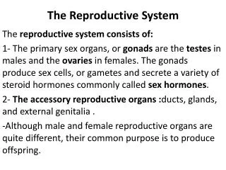
Hormones Classic definition: Chemical products secreted into the blood that act on distant tissues, usually in a regulatory fashion. Hormones can be released from endocrine and non-endocrine organs eg., the heart Hormone Effects: Endocrine Paracrine Autocrine Intracrine
Gonadotropin Releasing Hormone (GnRH) • GnRH was discovered independently by Drs. Roger Guillemin and Andrew V. Schally • Nobel Prize in Medicine in 1977 for their independent work that led to the isolation of hormones from the brain region known as the hypothalamus • Mammalian GnRH, termed GnRH-I: (Glu-His-Trp-Ser-Tyr-Gly-Leu-Arg-Pro-Gly) is a key regulator of the reproductive axis in diverse vertebrates. • A second GnRH subtype, termed GnRH-II, originally identified from chicken hypothalamus has been found in humans. • This second GnRH form differs from GnRH-I by three amino acid residues at positions 5, 7, and 8 (His5Trp7Tyr8GnRH-I).
The Hypothalamus The hypothalamus is located at the base of the forebrain and regulates homeostatis: metabolic/autonomic activities such as food intake, energy expenditure, body weight, fluid intake and balance, thirst, blood pressure, body temperature, sleep cycle, and reproduction.
The location of GnRH neurons is species-dependent Primates: in preoptic area, anterior hypothalamic area, and the medial basal Rodents: only in rostral areas- the preoptic area, and anterior hypothalamus GnRH neurons are relatively few in number, diffusely distributed in the POA and are not organized into nuclei GnRH neurons in the mouse and rat
The Pituitary Gland The pituitary gland, or hypophysis, the size of a pea sits in a small, bony cavity (sella turcica) at the base of the brain and is functionally connected to the hypothalamus Hypothalamus releases hormones into the pituitary (including GnRH) The posterior pituitary lobe (neurohypophysis)is directly connected to the brain and is derived from the neural ectoderm. The anterior (adenohypophysis) and intermediate lobes are derived from the oral ectoderm (FYI: In humans the intermediate lobe is a thin layer of cells between the anterior and posterior lobes)
The Hypothalamus-Pituitary Hypothalamic neuroendocrine neurons are comprised of two types of neurons that mediate hypothalamic endocrine functions: Magnocellular and Parvicellular Neurons Parvicellular neurons(hypophysiotropic cells) extend into the ME where they release their neuropeptides* into the anterior pituitary, via the portal circulation, where they increase or decrease synthesis and release of other hormones from the anterior pituitary into the general circulation (example: GnRH) Magnocellular neurons (neurohypophyseal cells), whose axons traverse the ME, extend directly into the posterior pituitary and release their neuropeptides* that are then released directly into the general circulation. *oxytocin and vasopressin **gonadotropin releasing hormone (GnRH) corticotropin releasing hormone (CRH or CRF) thyroid stimulating hormone releasing hormone (TRH) growth hormone releasing hormone (GHRH) somatostatin / growth hormone inhibiting hormone (GHIH)
Magnocellular neurosecretory neurons Neurohypophysis Posterior pituitary Adenohypophysis Anterior pituitary
Hypothalamus-Pituitary The hypothalamus releases hormones such as GnRH into the anterior pituitary via a bridgelike structure, the median eminence (ME). ME is one of seven areas of the brain devoid of a blood-brain barrier (BBB) and where axon terminals of hypothalamic neurons release (hypophysiotropic) neuropeptides involved in the control of anterior pituitary function. The ME is also traversed by the axons of hypothalamic neurons ending in the posterior pituitary Anterior Pituitary
Structure of the median eminence Figure 6 GnRH neurons GnRH neurons
Hypothalamus-Pituitary The Hypophysial Portal Vascular System • Hypophysiotropic peptides (eg, GnRH) released into the ME from hypothalamic nerve endings • They enter the 1o plexus capillaries (which receives blood from the carotid artery) • Transported to anterior pituitary via hypophysial portal veins (long parallel portal veins connect two capillary beds) located in the pituitary stalk • into the 2o plexus which supplies blood to the anterior pituitary • peptides exit the 2o plexus and act on receptors (eg, GnRH-R) on anterior pituitary cells regulating hormone production/secretion • These hormones then enter the same capillaries and are carried into the general circulation. Luteinizing Hormone and Follicle Stimulating Hormone (LH and FSH)
HYPOTHALAMIC-PITUITARY-GONADAL AXIS Feedback by Steroid Hormones: Estrogen Produced by granulosa cells/developing follicles Progesterone secreted from the developing corpora lutea (Testosterone) Negative or Positive Feedback Hypothalamus GnRH Anterior Pituitary FSH & LH Gonads
GnRH Feedback Regulation 1 LH pulse/60 mins 1 LH pulse/90 mins
Early in Follicular Phase: Estradiol is secreted by developing follicles, therefore estradiol is low. Correspondingly, there is a weak estradiol-induced inhibition of the GnRH pulse generator and LH pulse frequency is relatively fast at 1 pulse/60 mins. 1 LH pulse/60 mins 1 LH pulse/90 mins
Late Follicular Phase: As estradiol level builds up as follicular phase progresses, a stronger negative estradiol-induced regulatory feedback on the GnRH pulse generator is observed leading to a reduced LH pulse frequency of 1 pulse/90 mins 1 LH pulse/90 mins
Preovulatory GnRH/LH-Surge: However, as more estradiol is produced (see pre-ovulatory peak), a level is achieved that leads to a positive estradiol-induced feedback on the GnRH pulse generator and the surge release of LH and FSH and ovum release
The luteal phase: the empty follicle transformed into the corpus luteum. This becomes a rich source of progesterone (and some estradiol). This maintains pregnancy and together strongly negatively feeds back on the GnRH pulse generator.
Species Differences Rodents lack long luteal phase: Progesterone has positive feedback on preovulatory surge Primate and Sheep similar to Human
Steroid Feedback on GnRH neurons • GnRH neurons lack receptors for estradiol (ER alpha) and progesterone; • some contain ER beta, but this receptor is not involved with estradiol-feedback on GnRH neurons • So, steroids provide feedback on GnRH system indirectly via interneurons
Regulation of GnRH neurons • Much research is ongoing to further delineate the functional network that controls GnRH neurons • Numerous neuro- transmitters and –peptides have been identified to regulate GnRH (>35) • Kisspeptin is a major modulator
Steroids act during different times of life • Steroid feedback of GnRH secretion in adulthood: • Activational effects • Steroids act on GnRH system during puberty • Steroids act during early/prenatal life • Organizational effects: cause permanent changes to reproductive (and other systems)
Protein product: Testis-determining factor External genital tubercle Sexual differentiation of gonads
SEXUAL DIMORPHISM Prenatal steroids play critical role in sexual differentiation of brain and behavior A wide range of animal and human behaviors are sexually dimorphic (dimorphic means “two forms”). Some of these behaviors are reflexive in nature (reproduction), others require a high level of cognitive activity (spatial thinking, language).
DIFFERENTIATION OF THE BRAIN:THE ACTIVE AGENT IS ESTRADIOL • Testosterone is converted to estradiol once it has entered the relevant neurons (enzyme that converts testosterone to estradiol: aromatase). • Thus, it is estradiol/estrogen that acts inside neurons to stimulate sexually dimorphic patterns of neuronal circuitry as a result of the presence of testosterone in developing males. • Organizational effects of estradiol • Humans: prenatal • Rodents: perinatal (first week after birth)
TROPHIC EFFECT OF ESTROGEN(DOMINIQUE TORAN-ALLERAND, 1978) No estrogen Estrogen
ESTROGEN: TROPHIC FACTOR • Estradiol can produce brain dimorphisms by: • increasing neuronal size • nuclear volume (size of brain area) • dendritic length • dendritic branching • spine density • and number of synapses
EXAMPLES OF SEXUALLY DIMORPHIC BRAIN AREAS:HYPOTHALAMUS the sexual dimorphic nucleus (SDN; Roger Gorski and coworkers, 1978) SDN is small in females and large in males, and its development is under the influence of estradiol.
RAT SDN Male Female Female + Testosterone Female + Estrogen
Since then, other sexual dimorphic nuclei in hypothalamus have been described, as well as in amygdala, brainstem, spinal cord, cortex. Generally, these nuclei are larger in males than in females.
FUNCTION OF BRAIN SEXUAL DIMORPHISM? • Sexually dimorphism in reproductive function: • Female: ovulation • Reproductive behavior: • Female: feminine sex behavior; maternal behavior • Male: masculine sex behavior, requires defeminization and masculinization of behavior; aggression • Cognition and language • Disease onset and progression
Cell-autonomous action of sex-chromosome genes • Recent studies in rodents and birds have demonstrated that sexual differentiation can occur independent of gonadal hormones and involve sex chromosome genes • gynandromorph
Zebra finch gynandromorph Female ZZ Male ZW
Thank you Have lots of fun this week!