Download
1 / 83
1k likes | 1.85k Vues
Anatomy, Radiographic Evaluation, and Classification of Pelvic Ring Injuries. Robert M. Harris MD Medical Director of Orthopaedic Trauma Mountain States Health Alliance East Tenn State University Quillen School of Medicine Revised November 2010 Created March 2004 Revised April 2007

E N D
Anatomy, Radiographic Evaluation, and Classification of Pelvic Ring Injuries Robert M. Harris MD Medical Director of Orthopaedic Trauma Mountain States Health Alliance East Tenn State University Quillen School of Medicine Revised November 2010 Created March 2004Revised April 2007 By Kyle Dickson MD
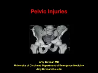
Pelvic Ring Disruption • Marker for severe injury • Overall mortality 6-10% • Life threatening
Magnitude of Forces • ACL injury 500-1000N • LC-I pelvic fracture 6000-9000N
Bone Anatomy • Two innominate bones with sacrum. • Coalesce at triradiate cartilage. • Ilium, ishium and pubis have three separate ossification centers that fuse at sixteen years. • Gap in symphysis < 5 mm • SI joint 2-4 mm
Ligamentous Anatomy • Ligaments - posterior ligaments are stronger than anterior ligaments: • Posterior SI • Anterior SI • Interosseous ligaments • Pubic symphysis • Sacrotuberous • Sacrospinous
ANATOMY Ligamentous ASI PSI ST SS ST
Posterior Ligaments • Ant. SI Joint – resist external rotation • Post. SI and Interosseous – posterior stability by tension band (strongest in body) • Iliolumbar ligaments augments posterior complex • Sacrotuberous (sacrum behind sacro-spinous into ischial tuberosily vertically)Resists shear and flexion of SI joint • Sacrospinous – (anterior sacral body to ischial spine horizontally) resists external rotation
Normal SI Joint Motion with Gait • < 6 mm of translation • < 6° rotation • Intact cadaver resist 5,837 N (1,212 lbs)
ANATOMY Relationships
Vascular Anatomy • Internal iliac artery courses medial to the vein, splits into anterior and posterior branches. • Posterior branch is more likely injured (SGA is largest branch). • Usual bleeding is from venous plexus.
Potentially Damaged Visceral Anatomy • Blunt vs. impaled by bony spike • Bladder/urethra • Rectum • Vagina
Pelvic Stability • Strength of ring: 40% anterior and 60% posterior. • Vsphere = 4/3r³. • Stability – ability of pelvic ring to withstand physiologic forces without abnormal deformation
IDENTIFY THE HIGH RISK PELVIC DISRUPTION By Radiography By Physical Exam
Physical Exam • Physical Exam-poor sensitivity (8%) for mechanically unstable pelvis fractures in blunt trauma patients • Shlamovitz GZ, Mower WR, Morgan MT-Journal of Trauma Mar 09
Radiographs • Anteroposterior (AP) • Inlet (40° caudad) • Outlet (40 ° cephalad) • CT scan • Judet (acetabular fractures)
AP VIEW If evidence of pelvic ring fracture...
Inlet (Caudad) View • Horizontal Plane Rotation • Posterior Displacement • Sacral ala
Outlet (Cephalad) View • Sacrum • Cephalad Displacement • Sacral Foramina
CT Scan • Better defines posterior injury • Amount of displacement versus impaction • Rotation of fragments • Amount of comminution • Assess neural foramina
Radiographic Signs of Instability • Sacroiliac displacement of 5 mm in any plane • Posterior fracture gap (rather than impaction) • Avulsion of fifth lumbar transverse process, lateral border of sacrum (sacrotuberous ligament), or ischial spine (sacrospinous ligament)
Translational Deformities • X axis – Diastasis or impaction • Y axis – Caudad or cephalad displacement • Z axis – Anterior or posterior displacement
Rotational Deformities • X axis – Flexion or extension • Y axis – Internal rotation or external rotation • Z axis – Abduction or adduction
Classification • Aids in predicting hemodynamic instability • Aids in predicting visceral and g.u. injuries • Aids in predicting pelvic instability • Aids in understanding mechanism of injury, force vector of injury, and surgical tactic for reduction
Classification Systems • Anatomical (Letournel) • Stability & Deformity (Pennal, Bucholz, Tile) • Vector force and associated injuries (Young & Burgess) • OTA-research
Anatomical Classification(Letournel) Where The Pelvis Breaks
Rami fractures Symphyseal disruption Iliac wing fracture Iliac wing/sacroiliac (SI) joint (crescent fracture) SI joint Sacrum/SI joint Sacrum fracture Anterior Posterior
Magnitude and direction of forces Lateral posterior compression (LC) Anterior posterior compression (APC) Vertical shear (VS) Added stability to the classification Pennal, 1961 Bucholz, 1981 Tile, 1988
Tile Classification • Type A: Stable fracture. • Type B: Rotationally unstable, but vertically stable. • Type C: Rotationally and vertically unstable.
OTA/AO – Pelvic Injury Classification • 61A – Lesion sparing (or with no displacement of ) posterior arch • B – Incomplete disruption at posterior arch; partially stable • C – Complete disruption of posterior arch; unstable
A Fractures – Ring Intact • A-1 – Fracture of innominate bone; avulsion • A-2 – Fracture of innominate bone; direct blow • A-3 – Transverse fracture of sacrum and coccyx
B-Ring Injury – Partially stable • B-1 – Unilateral partial disruption of posterior arch, external rotation (“open book” injury) • B-2 – Unilateral, partial disruption of posterior arch, internal rotation (lateral compression injury) • B-3 – Bilateral, partial lesion of posterior arch
C – Complete Disruption Posterior Arch, Unstable Pelvis • C-1 – Unilateral, complete disruption of posterior arch • C-2 – Bilateral, ipsilateral complete, contralateral incomplete • C –3 – Bilateral, complete disruption
Young-Burgess Radiology 1986 • Based on mechanism of injury • Predictive of associated local & distant injury • Useful for planning acute treatment
MECHANISM OF INJURY (MOI) • Do initial radiographs agree with MOI in pelvic ring disruptions- Linnau KF, Blackmore CC, Routt ML, Mock CN-J Ortho Trauma Jul 2007 • more reliable for LC than AP mechanisms
MECHANISM OF INJURY • Lateral compression(implosion) • AP compression(external rotation) • Vertical shear • Combined injury
Young-Burgess Classification • LATERAL COMPRESSION fracture of anterior ring plus: • LC -I Compression fracture of anterior sacrum • LC -II Iliac wing fracture posteriorly (unstable) • LC -III Windswept pelvis (contralateral SI injury) • ANTERIOR-POSTERIOR COMPRESSION • APC - I Partial disruption • APC - II Posterior sacroiliac ligaments intact • APC - III Posterior sacroiliac ligaments disrupted • VERTICAL SHEAR cephlad and posterior displacement • COMBINED MECHANISM (LC & VS most common)
CLASSIFICATION Mechanism and direction of injury
DISRUPTED PELVIC RING • Posterior/SI injury is a marker for associated vascular injuries • Tamponade efforts and fluid resuscitation may be rendered useless
Resuscitation • Young and Burgess classification: • LC III • APC II • APC III • VS • CM
RESUSCITATION REQUIREMENTS units blood 1st 24 hours
Mortality Deaths:
Interobserver Reliability of the Young/Burgess and Tile classifications • Koo H, Leveridge M, McKee,MD, Schemitsch EH, J Ortho Trauma Jul 2008 • Young/Burgess –Kappa .72-better for the training surgeon • CT-improved assessment of stability • Furey AJ, O”Toole RV, Turen C, Ortho June 2009 • Interobserver – moderate degree of agreement • Intraobserver- moderate for Tile • Substantial for Burgess
LATERAL COMPRESSION LC I: Sacral compression
Lateral Compression • Most common pattern. • LC1 – stable, load to posterior ring. • LC2 – load to anterior ring, posterior ligaments injured, ST and SS intact. • LC3 – LC2 + external rotation injury of the other side.
LATERAL COMPRESSION Common anterior pattern