Download
1 / 47
520 likes | 707 Vues
Learn about the epidemiology, anatomy, mechanisms of injury, and proper management of pelvic injuries, including essential work-up and clinical features. Essential exam components and diagnostic studies are highlighted, along with injury mechanisms and associated clinical findings.

E N D
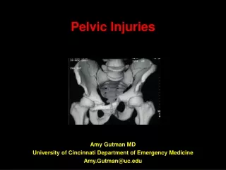
Pelvic Injuries Amy Gutman MD University of Cincinnati Department of Emergency Medicine Amy.Gutman@uc.edu
OUTLINE • Epidemiology • Anatomy • Mechanisms of Injury • Management • Conclusions
EPIDEMIOLOGY • 3-5% of all fractures • 60% vehicular trauma • 30% falls • 10% crush, athletic, penetrating trauma • Morbidity & Mortality • 6-19% mortality • 50% mortality with hemorrhagic shock • 58% mortality if age >65 • Children with proportionally greater hemorrhage • Associated abdominal, GU, chest, head trauma (early & late mortality) • Potential for severe hemorrhage (early mortality)
Support & hematopoeisis • Ilium, ischium, pubis, sacrum, coccyx • Stable five jointed ring • Lumbosacral, sacroiliac, sacro-coccygeal & symphysis pubis • Ring stability dependant upon posterior sacroiliac, sacro-tuberous & sacrospinous ligaments • Arcuate/ iliopectineal line divides false & true pelvis • Acetabulum: • Inner wall (pubis) easily fractured • Superior iliac bears weight • Posterior wall thick ischium
Left Pelvis (Outside View) Iliac Crest PSIS ASIS Ilium Post Semilunar Notch Ant Semilunar Notch Greater Sciatic Notch PIIS AIIS IschialSpine Lunate Face Ischium Ischial Tuberosity Pubic Spine Pubis Obturator Foramen Ramus
Iliac Crest ASIS AIIS Acetabulum Ischial Spine Sacrum Sacroiliac Joint Obturator Foramen Ischial Tuberosity Pubic Rami Female & Male Pelvic Anatomy
ESSENTIAL WORK-UP: History & Physical Examination Imaging Laboratory
MULTI-TASKING PELVIC TRAUMA • Simultaneous evaluation & management • ATLS • Trauma Labs • Secondary Survey: • Rectal & bimanual pelvic • Look for associated injuries, hemorrhage (+RP) • +/- reduce pelvic volume • Aggressive fluid resuscitation • Consult: • Orthopedics: external fixation • Interventional Radiology: 2% require embolization
ESSENTIAL EXAM COMPONENTS • Observation (joint, joint above, joint below): • Contour, height/ length discrepancies, open wounds, ecchymosis • Blood in foley • Auscultation/ Percussion: • Bowel sounds, bladder integrity (FAST US) • Palpation: • Pain, subq air, rebound, step-off on pelvic/ rectal examination • Compression: • Stability, ability to weight-bear • ROM: • Flex, extend, rotational; limited if spinal packaged
CLINICAL FEATURES • Correlation with Fracture: • Pain (55%) • Crepitus (95%) • Palpable instability (98%) • Other: • Perianal or pelvic edema • Ecchymosis • Lacerations • Deformity • Hematoma over inguinal ligament • Destot’s scrotal hematoma • Earle’s Sign (palpation of fracture w/ rectal examination)
Plain Radiograph • AP: standard • Inlet: AP displacement, post arch • Outlet: superior-inferior displacement sacrum • Judet: acetabular view • CT • Posterior arch • Acetabulum • RP hemorrhage • Associated injuries
Angiography • Diagnostic & therapeutic • FAST/ Bedside US • Less costly, quick diagnosis, statistically as accurate as CT for intra-abd injury • Supraumbilical DPL if no CT or US • Poor for RP bleed • RUG & RUC • Fractures with blood @ meatus, macroscopic hematuria, inability to void, perineal hematoma
Standing Support: • Femorosacral arch (arcuate line) • Tie arch (pubic bones & superior rami) • Seated Weight Bearing Forces: • Tie arch fractures first at symphysis, rami & lateral to SI joints • Large force required to disrupt bony pelvis: • 6+ liters blood into pelvis from post venous plexus • Nerve supply from lumbosacral plexuses • Lower urinary tract, bladder, peritoneal reflection, descending colon, sigmoid, rectum, anus, uterus, vagina
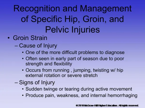
FRACTURE MECHANISM OF INJURY CLINIAL FINDINGS/ INJURIES* Iliac Wing Direct lateral trauma Swelling, tender iliac wing Abd pain, ileus, acetablular fxs Pubis/ Ramus Fall, direct trauma, exercise-induced Pain w/ ambulation, rectal injury stress fx, pregnancy Ischium Body Violent, fall from seated posit. Pain w/ mvt hamstring, rectal injury Least common Sacral AP trauma or fall in flexion, Pain w/ rectal exam. Sacral root transverse fractures injury w/ upper transverse Coccyx Fall in sitting position, common Pain over sacrum or w/rectal exam in women ASIS Sartorius contraction Pain w/ hip flexion/ abduction (hurdlers/ sprinters) AIIS Rectus femoris contraction Pain in groin or hip flexion (soccer) Ischial Tuberosity Hamstring contraction Pain with sitting, thigh flexion *Tintinelli
Iliac Wing Direct lateral trauma Swelling, tender iliac wing Abd pain, ileus, acetablular fxs
Pubis/ Ramus Fall, direct trauma, exercise- induced stress fractures, pregnancy Pain w/ ambulation Associated rectal injury
Coccyx Fall in sitting position Common in women Pain over sacrum or w/ rectal exam
ASIS Sartorius contraction Pain w/ hip flexion or abduction Hurdlers/ sprinters
AIIS Rectus femoris contraction Pain in groin or hip flexion Soccer
Ischial Tuberosity Hamstring contraction Pain with sitting, thigh flexion
TILE A “STABLE” • Fractures possibly meeting discharge criteria • A1: Innominate avulsion (ischial tuberosity/ iliac crest) • A2-1: Iliac wing (Duverney’s) • A2-2: Isolated rami • A2-3: “Straddle”; 4 pillar anterior ring, BL pubic rami • A3: Transverse sacrum or coccyx
YOUNG’S INJURY CLASSIFICATION: LATERAL COMPRESSION • Most common • 13% overall mortality • Broadsided MVC, pedestrian struck on side • Ipsi- or contralateral force causing transverse fracture
LATERAL COMPRESSION • I: Ipsilateral sacral compression • II: Ipsilateral iliac wing/ crescent fx • III: “Open Book”; lat compression w/ contralateral ant-post compression. 60% mortality
YOUNG’S INJURY CLASSIFICATION: ANTERIOR-POSTERIOR COMPRESSION • 25% all pelvic fractures • Head-on collisions • Posterior hip dislocations, symphyseal diastasis &/ or longitudinal rami fxs
ANTERIOR POSTERIOR • Diastasis &/ or longitudinal rami fx • Wide symphysis or ant sacro-iliac • Intact but stretched ligaments • 25% mortality
ANTERIOR POSTERIOR • II: Wide ant SI joint, disrupted ant sacroiliac, sacrotuberous, sacrospinous ligaments, intact post SI ligs. 28% mortality • III: Complete anterior & posterior SI disruption w/ lateral displacement. 53% mortality
APC: OPEN BOOK • Lateral + ant + posterior compression • If unilateral: vertically stable, rotationally unstable • Posterior SI ligaments intact providing vertical stability • Symphysis >2.5cm = injury to posterior sacroiliac ligaments • Associated Injuries: • Posterior arch & SI disruption • NV injury • RP hematoma expansion • Bladder rupture, urethral & pelvic vascular injury
APC: BUCKET HANDLE • Posterior + lateral + upward rotary force • Ipsilateral sacroiliac disruption w/ contra-lateral pubic rami fracture • Ipsilateral hemipelvis rotated superior-medially
YOUNG’S INJURY CLASSIFICATION: VERTICAL SHEAR • 5% all pelvic fractures • Fall from significant height • Fracture fragments vertically aligned • Vertical displacement anteriorly & posteriorly through sacroiliac joint, iliac wing or sacrum
VERTICAL SHEAR • Vertical displacement anteriorly & posteriorly through SI, iliac wing &/ or sacrum • 75% massive hemorrhage • 25% mortality
MALGAIGNE VS • Least common • Fall onto fixed lower extremities • Longitudinal fractures through anterior & posterior pelvis • Symphysis disrupted > 3-4 cm, rami & post displacement with unstable • sacrum & SI joint • >75% mortality from vascular, abdominal & thoracic injuries
YOUNG’S INJURY PATTERNS: COMBINATION • Commonly lateral compression & vertical shear • 58% mortality
CHIH/PTXSpleen Liver RP Bladder UreterMort LCI 61 11 5 6 4 14 3 LC292 33 25 0 8 25 8 7 LC3 75 33 20 0 3 27 7 APC272 0 11 0 0 11 6 20 APC3 71 14 14 14 28 43 0 VS56 33 11 0 22 0 0 <1 Mixed73 0 0 9 9 18 9 18 J Trauma 1990; 30:848 Non-Ortho Injuries Assoc w/ Young Pelvic Fxs
ATLS • Stabilize • Evaluate for associated injuries • With the exception of ligamentous injuries in otherwise healthy adults resulting from minor trauma, all patients should be admitted to orthopedics or trauma services • Possible IR, urology, general surgery, nephrology, & / or neurosurgery consultations
Biffl WL, et al. Ann Surg. 2001 Jun;233(6):843-50. Evolution of a multi-disciplinary clinical pathway for the management of unstable patients with pelvic fractures • Pathway I:*Pathway II: • Trauma team/ surgeon in ED *Orthopedic Surgeon • Early blood transfusions • Prompt RX of associated injuries • Pelvic stabilization • Early pelvic angiography & embolization • Findings: • Pelvic binding replaced traditional ex-fix devices • US/ CT scan replaced DPL • Deaths 31% to 15%, death within 24 hrs 16% to 5%, death from exsanguination 9% to 1%, death from MOF 12-1% • “Evolution of a multidisciplinary clinical pathway, coordinating resources of a level I trauma center, directed by joint decision making b/w trauma surgeons & traumatologists resulted in improved survival. Primary benefits… reducing early deaths from exsanguination & late deaths from multiple organ failure”
PELVIC BINDERS • Tamponade active bleeding • Minimize cavity available to fill with blood • No difference between binders
External fixation complications from initial poor bleeding control • Unstable fractures to angiography if no laparotomy • If laparotomy indicated, external fixation placed intra-operatively followed by post-operative angiography
SURGERY • Early mortality secondary to uncontrolled hemorrhage • Late mortality due to sepsis/ MOF • Limited surgical intervention reduces risk of systemic complications/ late mortality • Unless laparotomy required, hemodynamic instability from unstable pelvic fractures best approached with a combination of emergency external fixation hemostasis & pelvic tamponade
Never underestimate the force required to injure a pelvis • Always expect a second (or 3rd, 4th…) injury • Mechanism may provide a diagnosis before examination • Surgery is never first line for an isolated pelvic injury