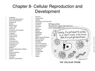
Chapter 27 Reproduction and Embryonic Development
680 likes | 862 Vues
0. Chapter 27 Reproduction and Embryonic Development. 0. Baby Bonanza The increased use of fertility drugs Has caused an increase in the number of multiple births in the United States. 0. Fertility drugs Are sometimes too effective. ASEXUAL AND SEXUAL REPRODUCTION. 0.

Chapter 27 Reproduction and Embryonic Development
E N D
Presentation Transcript
0 • Chapter 27 • Reproduction and Embryonic Development
0 • Baby Bonanza • The increased use of fertility drugs • Has caused an increase in the number of multiple births in the United States
0 • Fertility drugs • Are sometimes too effective
ASEXUAL AND SEXUAL REPRODUCTION 0 • 27.1 Sexual and asexual reproduction are both common among animals • In asexual reproduction • One parent produces genetically identical offspring
0 • Asexual reproduction • Enables an individual to produce many offspring rapidly
Eggs 0 • Reproduction in some animals is accomplished through external fertilization • In which the parents release their gametes into the environment where fertilization occurs Figure 27.1E
0 • Sexual reproduction • May enhance reproductive success in changing environments
HUMAN REPRODUCTION 0 • 27.2 Reproductive anatomy of the human female • Both sexes in humans • Have a set of gonads where gametes are produced • Have ducts for delivery of the gonads and structures for copulation
Ovaries Oviduct Follicles Corpus luteum Wall of uterus Uterus Endometrium (lining of uterus) Cervix (“neck” of uterus) Vagina 0 • A woman’s ovaries • Contain follicles that nurture eggs and produce sex hormones Figure 27.2A
Egg cell Ovary LM 200 0 • Oviducts convey eggs to the uterus • Where the eggs develop Figure 27.2B
0 • The uterus opens into the vagina • Which receives the penis during intercourse and forms the birth canal
Oviduct Ovary Uterus Rectum (digestive system) Urinary bladder (excretory system) Pubic bone Cervix Urethra(excretory system) Vagina Shaft Glans Clitoris Bartholin’s gland Prepuce Labia minora Anus (digestive system) Labia majora Vaginal opening 0 • Other structures of the female reproductive system include • Labia, the clitoris, Bartholin’s glands, and the cervix Figure 27.2C
Rectum (digestive system) Urinary bladder (excretory system) Seminal vesicle Vas deferens Ejaculatory duct Pubic bone Erectile tissue of penis Prostate gland Bulbourethral gland Urethra (excretory system) Penis Vas deferens Epididymis Glans of penis Testis Prepuce Scrotum 0 • 27.3 Reproductive anatomy of the human male • A man’s testes • Produce sperm Figure 27.3A
Urinary bladder (excretory system) Seminal vesicle (behind bladder) Prostate gland Bulbourethral gland Urethra Erectile tissue of penis Scrotum Vas deferens Epididymis Testis Glans of penis 0 • Several glands • Contribute to the formation of fluid that nourishes and protects sperm Figure 27.3B
Urethra region here expands and fills with semen Urinary bladder Sphincter contracts Contractions of vas deferens Contractions of seminal vesicle Contractions of prostate gland Contractions of epididymis Sphincter contracts First stage Semen expelled Sphincter remains contracted Contractions of muscles around base of penis Contractions of urethra Sphincter relaxes Second stage 0 • During ejaculation • Sperm and the nourishing fluid, called semen, are expelled through the penis Figure 27.3C
Stimuli from other areas in the brain Hypothalamus Releasing hormone Anterior pituitary Negative feedback LH FSH Androgenproduction Testis Sperm production 0 • A negative feedback system of hormones • Controls sperm production Figure 27.3D
Epididymis Penis Testis Scrotum 2n Diploid cell Testis Seminiferous tubule Differentiation andonset of Meiosis I Cross section ofseminiferoustubule 2n Primary spermatocyte (in prophase of Meiosis I) Meiosis I completed Secondary spermatocyte n n (haploid; double chromatids) Meiosis II Developing sperm cells(haploid; single chromatids) n n n n Differentiation Sperm cells n n n n Center ofseminiferous tubule (haploid) 0 • Primary spermatocytes, which are diploid, are made continuously in the testes • And undergo meiosis to produce haploid sperm Figure 27.4A
In embryo Diploid cell 2n Differentiation andonset of Meiosis I Primary oocyte 2n Present at birth (arrested in prophaseof Meiosis I) Completion of Meiosis Iand onset of Meiosis II Secondary oocyte n Firstpolar body (arrested at meta-phase of Meiosis II;released from ovary) n Entry of sperm triggerscompletion of Meiosis II Ovum Secondpolar body n n (haploid) 0 • Each month one primary oocyte • Matures to form a secondary oocyte, which can be fertilized • Completes meiosis and becomes a haploid ovum Figure 27.4B
Degeneratingcorpus luteum Start: Primary oocyte within follicle Corpus luteum Growingfollicles Mature follicle Ovary Secondaryoocyte Ruptured follicle Ovulation 0 • The development of an ovarian follicle • Involves many different processes Figure 27.4C
0 • 27.5 Hormones synchronize cyclic changes in the ovary and uterus • The ovarian cycle includes • Changes in the ovary that occur about every 28 days • The menstrual cycle • Involves changes that occur in the uterus
0 • An Overview of the Ovarian and Menstrual Cycles • Events in the menstrual cycle • Are synchronized with the ovarian cycle, which occurs about every 28 days
0 • Uterine bleeding, called menstruation • Includes the breakdown of the endometrial lining • Usually persists for 3–5 days • After menstruation • The endometrium, the lining of the uterus, regrows
0 • Five hormones • Synchronize the events in the ovarian cycle Table 27.5
0 • Hormonal Events Before Ovulation • Approximately every 28 days • The hypothalamus signals the anterior pituitary to secrete FSH and LH • FSH and LH • Trigger the growth of a follicle
0 • As the follicle grows, it secretes estrogen • Which causes a burst in FSH and LH levels, leading to ovulation
0 • Hormonal Events at Ovulation and After • After ovulation • The follicle becomes the corpus luteum • The corpus luteum secretes both estrogen and progesterone • Which exert negative feedback on the hypothalamus and pituitary, causing a decline in FSH and LH levels
0 • As FSH and LH levels drop • The hypothalamus can once again stimulate the pituitary to secrete more FSH and LH, and a new cycle begins
0 • Control of the Menstrual Cycle • The menstrual cycle • Is directly controlled by estrogen and progesterone
0 • If fertilization of an egg occurs • A hormone from the embryo maintains the uterine lining and prevents menstruation
The ovarian and menstrual cycles A Control by hypothalamus Inhibited by combination of estrogen and progesterone Stimulated by high levels of estrogen Hypothalamus Releasing hormone Anterior pituitary FSH LH 1 B Pituitary hormones in blood 4 LH peak triggers ovulation and corpus luteum formation 6 LH FSH FSH LH 2 Ovarian cycle C 5 Corpus luteum Degenerating corpus luteum Growing follicle Mature follicle Ovulation Pre-ovulatory phase Post-ovulatory phase Progesterone and estrogen Estrogen D Ovarian hormones in blood 3 7 8 Estrogen Progesterone Progesterone and estrogen Estrogen Menstrual cycle E Endometrium 0 5 10 14 15 20 25 28 Days Menstruation Figure 27.5
0 • 27.6 The human sexual response occurs in four phases • The excitement phase • Prepares the sexual organs for coitus • The plateau phase • Is marked by increases in breathing and heart rate
0 • Orgasm follows • Characterized by rhythmic contractions of the reproductive structures • The resolution phase • Completes the cycle and reverses the previous responses
CONNECTION 0 • 27.7 Sexual activity can transmit disease • Sexual intercourse • Carries the risk of exposure to sexually transmitted diseases (STDs)
0 • STDs common in the United States Table 27.7
CONNECTION 0 • 27.8 Contraception can prevent unwanted pregnancy • Contraception • Is the deliberate prevention of pregnancy Table 27.8
Skin patch Condom Diaphragm Spermicide Birth control pills 0 • Contraception can be accomplished • Through various methods Figure 27.8
PRINCIPLES OF EMBRYONIC DEVELOPMENT 0 • 27.9 Fertilization results in a zygote and triggers embryonic development • Embryonic development begins with fertilization • The union of sperm and egg to form a diploid zygote
Colorized SEM 500 0 • The Properties of Sperm Cells • Only one sperm • Fertilizes an egg Figure 27.9A
Middlepiece Plasma membrane Neck Head Tail Mitochondrion(spiral shape) Nucleus Acrosome 0 • During fertilization • A sperm releases enzymes from the acrosome that pierce the egg’s coat Figure 27.9B
0 • The Process of Fertilization • Sperm surface proteins bind to egg receptor proteins • Sperm and egg plasma membranes fuse, and the two nuclei unite
0 • Changes in the egg membrane • Prevent entry of additional sperm • The fertilized egg (zygote) • Develops into an embryo
1 The spermapproachesthe egg The sperm’sacrosomal enzymesdigest the egg’s jellycoat 2 3 Proteins on thesperm head bind toegg receptors The plasmamembranes of spermand egg fuse 4 The sperm nucleusenters the eggcytoplasm 5 A fertilizationenvelope forms 6 The nuclei of sperm and egg fuse 7 • The process of fertilization Sperm Nucleus Spermhead Acrosome Plasmamembrane Acrosomalenzymes Receptor proteinmolecules Plasmamembrane Spermnucleus Vitellinelayer Cytoplasm Eggnucleus Jellycoat Egg cell Zygote nucleus Figure 27.9C
Zygote 2 cells 4 cells 8 cells Blastocoel Many cells(solid ball) Blastula(hollow ball) Cross sectionof blastula 0 • 27.10 Cleavage produces a ball of cells from the zygote • Cleavage is a rapid series of cell divisions • That results in a blastula, a ball of cells Figure 27.10
Ectoderm 0 • 27.13 Changes in cell shape, cell migration, and programmed cell death give form to the developing animal • Cells of the ectoderm • Fold inward during neural tube formation Figure 27.13A
Apoptosis Dead cellengulfed anddigested byadjacent cell 0 • Programmed cell death, or apoptosis • Is a key developmental process in which cells die Figure 27.13B
0 • 27.14 Embryonic induction initiates organ formation • In a process called induction • Adjacent cells and cell layers influence each other’s differentiation via chemical signals
Lens ectoderm Optic cup Cornea Futurebrain Lens Opticvesicle Futureretina Opticstalk Figure 27.14 • Induction during eye development 1 2 3 4
0 • 27.15 Pattern formation organizes the animal body • Pattern formation • Is the emergence of the parts of a structure in their correct relative positions
HUMAN DEVELOPMENT 0 • 27.16 The embryo and placenta take shape during the first month of pregnancy • Pregnancy, or gestation • Is the carrying of developing young within the female reproductive tract
Cleavage starts Fertilizationof ovum Ovary Oviduct Secondaryoocyte Blastocyst(implanted) Ovulation Endometrium Uterus 0 • An Overview of Developmental Events • Human development • Begins with fertilization in the oviduct Figure 27.16A