Download
1 / 40
560 likes | 1.63k Vues
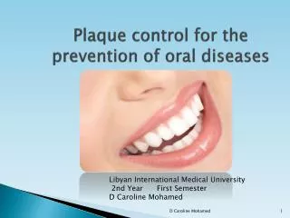
Plaque control for the prevention of oral diseases. Libyan International Medical University 2nd Year First Semester D Caroline Piske de A. Mohamed . Objectives. At the end of this topic you should be able to explain and describe: Plaque retentive factors.

E N D
Plaque control for the prevention of oral diseases Libyan International Medical University 2nd Year First Semester D Caroline Piske de A. Mohamed
Objectives • At the end of this topic you should be able to explain and describe: • Plaque retentive factors.
1. IATROGENIC FACTORS • Inadequate dental procedures that contribute to the deterioration of the periodontal tissues are referred as iatrogenic factors. • Margins of restoration • Contours and open contact • Materials • Design of RFD
a) Margins of restoration • Overhanging margins of dental restoration contribute to the development of periodontal disease: • Shift on gingival sulcus flora • No access to remove plaque Subgingival margins = large amount of plaque = more severe gingivites and deep pocket
b) Contours and open contacts • Over contoured crowns and restoration tend to accumulate plaque and possibly prevent the selfcleaning mechanism of the cheek, lips and tongue. • Contour of the occlusal surface as stablished by marginal ridges and related developmental grooves serves to deflect food away from the interproximal spaces. • The integrity and location of the proximal contacts along with the contour of the marginal ridges and developmental grooves prevent interproximal food impactation.
As the teeth wear down their originally convex proximally surfaces become flattened and the wedging effect of the opposing cusp is exaggerated. ( plunger cusps) • Not replaced missing teeth may also act as plunger cusps as the relationship between proximal contacts is altered. • The presence of abnormalities does not necessarily lead to food impaction and periodontal disease.
Ecxessive anterior overbite is a common cause of food impaction on the lingual surfacs of the opposing mandibular teeth.
c) Materials • Plaque that forms at the margins of the restoration ( all types of restorative material – silicate) is similar to that found on the adjacent non restored tooth surfaces. • Hygiene Pontic gives access for oral hygiene.
d) Design of Removal Partial Dentures. • After the insertion of partial dentures, the mobility of the abutment teeth, gingival inflammation and periodontal pocket formation increases because partial dentures favor the accumulation of plaque particularly if they cover the gingival tissue. • Take off PRD at night.
2. Associated with clinical procedure malocclusion • A maloccusion is a irregular alignment of teeth that may make plaque control more difficult.
2 ASSOCIATED WITH CLINICAL PROCEDURES • Maloclusion and Periodontal complications associated with therapy • Extraction of impacted 3 rd molar • Radiation therapy
Periodontal complication associated with the orthodontic therapy • Ortho. Therapy may affect the periodontium by favoring the plaque retention or by directly injuring the gingiva as a result of overextended bands and by creating excessive force on tooth and supporting structures. • Ortho. Appliances can modify the gingival ecosystem.
Gingival trauma and alveolar bone height • Ortho. Treatment is often started soon after eruption of the permanent teeth, when junctional epithelium is still adherent to the enamel surface. • Ortho. Bands should not be forcefully placed beyond the level of attachment because this will detach the gingiva from the tooth and result in apical proliferation of the junctionalep., with increased incidence of gingiva recession. • It is important to avoid excessive force and too rapid movement in ortho. Treatment.
Associated with clinical procedures • Extraction of impacted third molars Numerous studies reported that the extraction of impacted 3rd molars often results in the creation of vertical defects distal to the second molars.
Associated with clinical procedures • Radiation therapy • Citotoxic effects on both normal cells and malignant cells. • Periodontal attachment loss and tooth loss were greater on the radiated side in cancer patients treated with high dose unilateral radiation compared with non radiated control side of dentition.
Radiation therapy induces • Obliterative end arteritesresult in soft tissue ischemia and fibrosis • Irradiated bone becomes hypo vascular and hypoxic • Saliva production is permanentely impaired • Xerostomia results in greater plaque accumulation and a reduced buffering capacity from the remaining saliva
3. HABITS AND SELF INFLICTED INJURIES • Toothbrush trauma • Chemical irritation • Mouth breathing • Tongue thrusting • Tobacco use • Others
3. Habits and self inflicted injuries • Toothbrush trauma • Abrasion of the gingiva as well as alterations in tooth structure may result from aggressive brushing in a horizontal or rotary fashion. • Highly abrasive dentifrice. • Scuffing of epithelial surface, denudation of underlying connective tissue – gingival ulcer gingival recession.
Improper use of dental floss may result in lacerations of interdental papilla. • Interproximalattachment loss is generally a consequence of bacteria induced periodontitis, where as buccal and linghal attachment loss is frequently result of toothbrush abrasion.
Tobacco use • Smoking is one of the most significant risk factors currently available to predict the development and progression of periodontitis. • A diminished response to non surgical therapy has been reported for smokers.
Habits and self inflicted injuries • Chemical irritation • Acute gingival inflammation may be caused by chemical irritation resulting from either sensitivity or non specific injury. • Chemicals- Strong mouthwashes, topical application of corrosive drugs as aspirin, and accidental contact with phenol or silver nitrate, bleaching.
Mouth Breathing • Can dehydrate the gingival tissue and increase suscetibility to inflammation. • These patients may or may not have increased levels of dental plaque. • Tongue thrust
Tongue thrusting is often associated with an anterior open bite. During swallowing tongue is thrusted forward against the teeth instead of being placed against the palate. • When the amount of pressure against the teeth is great it can lead to tooth mobility and cause increased spacing of ant. Teeth.
Habits and self inflicted injuries • Fingernail biting • Using toothpicks • Trauma associated with oral jewelry (tongue and lip piercing) • Trauma associated with drug abuse
4. Anatomic contributing factors • Proximal contact relation • Cervical enamel projections and enamel pearls • Intermediate bifurcation ridge • Root anatomy • Cemental tears • Accessory canals • Root proximity • Adjacent teeth
a) Proximal contact relation • Open interproximal contacts or uneven marginal ridge relations are factors that may predispose to food impaction. • It can lead to inflammation, bone los, and attachment loss. • Open interproximal contacts that are easily cleansable may be as healthy as those with a proper contact relation.
b) Cervical enamel projections and enamel pearls • CEP appear as narrow wedge shaped extensions of enamel pointing from the CEJ towards furcation area. • Most frequently in molars. • Plaque retentive and can predispose to furcation involvement.
c) Intermediate bifurcation ridge Bifurcation ridges are one of the contributing anatomical factors in the etiology and compromised prognosis of furcation involved teeth.
d) Root anatomy • Palatogingival groove • Attachment area • Root trunk lengh • Interroot separation • Root fusion
Root grooves are developmental anomalies in which an infolding of the inner enamel epithelium and Hertwigs epithelial root sheath (HERS) creates a groove on the tooth surface. • Such morphological features compromise patient's self care, favour accumulation of plaque, calculus and food debris. • They facilitate plaque growth and later provide anaerobic condition for bacterial selection and proliferation. They cause patients inaccessibility to routine oral hygiene procedures and they also complicate restorative procedures.
Root trunk length is defined as area of the tooth extending from CEJ to the furcation. Therefore horizontal attachment loss leading to furcation invasion compromises the root trunk, resulting in the loss of one third of the total periodontal support. • The significance of root trunk is related to both prognosis and treatment of tooth. • A molar with a short root trunk is more vulnerable to furcation involvement but has a better prognosis after treatment since less periodontal destruction has presumably occurred. • Alternatively a furcation involved molar with a long root trunk and short roots may not be a candidate for root resection, since these teeth lose more periodontal support with furcation invasion.
e) Cemental tears • A Cemental tear is a piece of detached cementum, often with some dentin, that may remain attached to periodontal ligamental fibers. • It can lead to rapid periodontal bone loss and produce bony defect.
f) Accessory canals • Accessory canals may furnish a communication between canal and the PDL. • Pulpal necrosis could contribute to formation of periodontal defect through an accessory canal.
g) Root proximity • Close approximation of tooth roots, with an accompanying thin interproximal septum, leads to an increased risk of periodontal destruction. • Crowns of these teeth especially anterior teeth are very closely approximated and may have long interproximal contacts, and minimal embrasure space, which makes plaque removal difficult.
h) Adjacent teeth • Retention of periodontally compromised tooth may have a detrimental effect on a adjacent periodontally healthy tooth. • Adjacent third molars are of particular concern in patients with periodontites
Recommended • Mouth Care for Patients Receiving Chemotherapy and Radiation Therapy In. http://www.cancerlearning.gov.au/docs/Mouth_cares.pdf
Activity • Please buy and use 3 different types of tooth pastes. • Next class you should provide a report comparing the advantages and disadvantages of the different characteristics of each tooth paste regarding taste, smell, appearance, ease of use, feeling of freshness after brushing, effectiveness, prices and general satisfaction with it.
References • Marya CM. A Textbook of Public Health Dentistry. JaypeenBrothers Medical Publishers, 2011. • Harris. N.O.Primary Preventive Dentistry. Ed.Apleton& lange. 4 ed. 1991. • Leknes KN. The influence of anatomic and iatrogenic root surface characteristics on bacterial and periodontal destruction: A Review. J periodontal 1997; 68(6) :507-516.2. Matthews DC, Tabesh M. Detection of localized tooth -related factors that predispose to periodontal infections. Periodontol 2000; 2004;34:136-503. Blieden T M. Tooth related issues. Ann periodontal 1999;4(1): 91-74. Buckley LA. The relationship between malocclusion and periodontal disease. J Periodontol 1972; 43(7): 415-7.5. Wasserman B H, Thompson RH Jr, Geiger AM, Goodman SF, Pomerantz J, Tyugeon LR, BeubeFE. Relationship of occlusion and periodontal disease. II. Periodontal status of study population. J Periodontol. 1971;42(6): 371-8.6. Silness J, Roynstrand T, Relationship between alignment conditions of teeth in anterior segments and dental health. J Clin Periodontol 1985; 12(4): 312-20.7. Kornman KS, Loe H. The role of local factors in the etiology of periodontal diseases. Periodontol 2000 1993 2; 83-978. Jernberg GR., Bakdash MB, Keenan KM. Relationship between proximal tooth open contacts and periodontal disease. J Periodontol 1983; 54(9): 529-33.9. Koral SM, Howell T.H, Jeffcoat MK. Alveolar bone loss due to open interproximal contacts in periodontal disease. J Periodontol 1981 ;52(8): 447-50.10. Geiger A M, Wasserman B H, Turgeon L R . Relationship of occlusion and periodontal disease. 8. - Relationship of crowding and spacing to periodontal destruction and gingival inflammation. J Periodontol 1974; 45(1):43-49.11. Woofler C. The prevalence and etiology of gingival recession. Periodontal Abstr. 1969; 17(2): 45-5012. Grant, Stern , Listgarten. Treatment of periodontal trauma. Periodontics , sixth edition. C.V Mosby company 1988.13. Kepic TJ, O'Leary TJ. Role of marginal ridge relationship as an etiologic factor in periodontal disease. J Periodontol 1978; 49(11): 570-5.14. Haney JM, Leknes KN, Lie T, Selvig KA, Wikesjo UM. Cemental tear related to rapid periodontal breakdown: A case report. J Periodontol 1992; 63(3):220-4.15. Ishikawa I, Oda S., Hayashi J., Arakawa S. Cervical cemental tears in older patients with adult Periodontitis. Case reports. J Periodontol 1996; 67(1) :15-20.16. Gher ME, Vernino AR. Root morphology - clinical significance in the pathogenesis and treatment of periodontal disease. J Am Dent Asoc 1980 ;101(4): 627-33.17. Gher MW Jr, Dunlap RW . Linear variation in root surface area of maxillary first molar. J Periodontol 1985; 56(1): 39-43.18. Mass E, Aharoni K, Vardimon AD. Labial-cervical-vertical groove in maxillary permanent incisors- prevalence, severity and affected soft tissue. Quintessence Int 2005 ;36(4) :281-6.19. AL-Shammari KF, Kazor CE, Wang HL.Molar root anatomy and management of furcation defects. J Clin Periodontol 2001:28(8):730-40.20. Leknes KN, Lie T, Selvig KA. Root grooves: A risk factor in periodontal attachment loss . J Periodontol 1994;65(9):859-63.21. Hou GL, Tsai CC. Relationship between palato-radicular grooves and localized periodontitis. J Periodontol 1993;20(9) :678-82.22. Hou GL, Chen YM, Tsai CC, Weisgold AS. A new classification of molar furcation involvement based on the root trunk and horizontal and vertical bone loss. Int J Periodontics Restorative Dent 1998; 18(3): 257-65.23. Svardstrom G, Wennstrom JL. Furcation topography of the maxillary and mandibular first molars. J Clin Periodontol 1988; 15(5) :271-5.24. Santana RB, Uzel MI, Gusman H, Gunaydin Y, Jones JA, Leone CW. Morphometric analysis of the furcation anatomy of mandibular molars. J Periodontol 2004; 75(6): 824-9.25. Hou GL, Tsai CC. Cervical enamel projection and intermediate bifurcational ridge correlated with molar furcation involvements. J Periodontol 1997; 68(7): 687-93.26. Swan RH, Hurt WC. Cervical enamel projections as an etiologic factor in furcation involvement . J Am Dent Assoc 1976; 93(2) :342-5.27. Bissada NF, Abdelmalek RG. Incidence of cervical enamel projections and its relationship to furcation involvement in Egyptian skull. J Periodontol 1973; 44(9) :583-5.28. Leib AM, Berdon JK. Furcation involvements correlated with enamel projections from the cementoenamel junction. J Periodontol 1967; 38(4): 330-4.29. Shiloh J, Kopezyk R. Developmental variations of tooth morphology and periodontal disease. J Am Dent Assoc 1979;99(4): 627-630.30. Bower RC. Furcation morphology relative to periodontal treatment. Furcation root surface morphology. J Periodontol 1979; 50(7):366-74.31. Booker BW 3rd, Loughlin DM. A morphologic study of the mesial root surface of the adolescent maxillary first bicuspid. J Periodontol 1985; 56(11) :666-70. • 1. Löe H, Schiött CR, Karring G, Karring T. Two years oral use of • chlorhexidine in man. I. General design and clinical effects. J Periodontal • Res. 1976 Jun;11(3):135-44.