H3 Absorption of digested foods
100 likes | 245 Vues
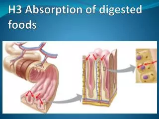
H3 Absorption of digested foods. Ms.TRS. H3.1 Structure of the ileum. The villi increase the surface area of the ileum for the absorption by ten times . The crypts contain the secretory cells of intestinal secretion. Mucosa is a mucus secreting membrane.

H3 Absorption of digested foods
E N D
Presentation Transcript
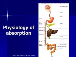
H3.1 Structure of the ileum • The villi increase the surface area of the ileum for the absorption by ten times. • The crypts contain the secretory cells of intestinal secretion. • Mucosa is a mucus secreting membrane. • The circular and longitudinal muscles combine to create the contractions known as peristalsis that maintains the movement of chyme along the alimentary canal. • The serosa is a tough outer membrane composed of collagen.
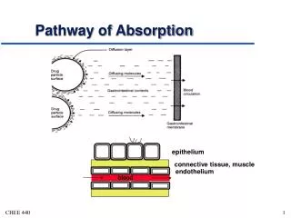
. H3.2 Ultrastructure of the epithelial gut cells • Epithelial cell: • Microvilli border increases the surface area for absorption of digested foods by many hundreds of times. • Tight Junctions in which adjacent cells closely join there plasma membranes to prevent leakage between the cells. • There is a large prominent nucleus in a medial to basal position. • There are many mitochondria reflecting the active transport of some absorbed molecules and the synthesis of lipoproteins along with other molecules. • Pinocytotic vesicles are very small vesicles formed by endocytosis. The purpose of these is to increase the surface area of membrane for other processes such as active transport or facilitated diffusion
H3.3 Absorption in the small intestine • Absorption of fatty acids . • (a) Bile salts form a micelle around fatty acids. The phospholipid structure of the salts allows it to fuse with the cell membrane and the fatty acid molecules to pass into the epithelial cells of small intestine villus. • b) The fatty acids and glycerol recombine in the endoplasmic reticulum to form lipid. • c) Protein is added to the lipid to form lipoprotein. This is how lipid is transported around the body. • d) The lipoprotein (called chylomicrons) is formed into small vesicles • e) Exocytosis of the vesicles releases the lipoprotein from the cell • f) The lipoprotein is taken up in the lacteal vessel a branch of the lymphatic system. • g) The lacteals, lymphatic system and the lipoproteins eventually enter the general circulation.
Facilitated diffusion: possible water soluble minerals and vitamins • Larger molecules move passively through the membrane via channel proteins • These proteins have large globular structures and complex 3d-shapes • The shapes provide a channel through the middle of the protein, the 'pore' • The channel 'shields' the diffusing molecule from the non-charged regions of the membrane.
Active Transport : Glucose absorptionand Amino acids absorption into the epithelial cells. • Active mean that the membrane protein 'pump' requires energy to function • This moves the molecules from low to high concentration against the concentration gradient • The energy causes a shape change in the protein that allows it to move the molecule to the other side of the membrane. • These membrane pumps are often closely associated with membrane immobilised enzymes.(H2.6)
. . • Endocytosis: probably pinocytosis • The formation of tiny vesicles by endocytosis is normally referred to as pinocytosis. • This increases the surface area for the processes of active transport and diffusion. • But in lipid absorption the micelle fuses with the cell membrane. The absorption of actual lipids would occur passively across the cell membrane
Cholesterol and the fat-soluble vitamins (A, D, E, K) are absorbed into the epithelial cells of the ileum by lipid diffusion Mineral ions and water-soluble vitamins are absorbed by passive transport in the ileum Dietary monosaccharides are absorbed by active transport in the ileum Water is absorbed by osmosis in the ileum and colon.
H3.4 Egestion Cellulose is an undigested insoluble polysaccharide which forms the fibre in the diet. Bile Pigments colour the egested faeces and are eliminated. The composition of these pigments often reflect liver function and are sometimes used as a diagnostic for that organs function. The gut retains its own bacterial populations with their own ecology. There is evidence to believe that normal digestion requires the presence of these populations and that factors that affect these populations (antibiotics) can lead to digestive disorders. Intestinal cells are constantly removed from the lining of the gut. Gut epithelium has a structure similar to that of skin, with which it shares a common embryological origin. Such cells are egested.