Download

1 / 21
210 likes | 245 Vues
This comprehensive guide explores osteoarthritis and rheumatoid arthritis, detailing types, clinical manifestations, pathologic changes, and pathogenesis. Learn about gout, tuberculous arthritis, and more.

E N D
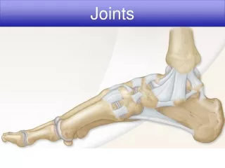
Osteoarthritis • Degenerative joint disease, is the most common disorder of the joints • Degeneration of the articular cartilage • The joints commonly involved : hips, knees, lower lumbar and cervical vertebrae, proximal and distal interphalangeal joints of the fingers.
Types • Primary • secondary • Previously deformed joint, • joint degeneration that occurs in the context of certain metabolic disorders, hemochromatosis, diabetes mellitus
Clinically • Most individuals with primary osteoarthritis are asymptomatic until after the age of 50 • joint stiffness and deep, aching pain, particularly in the morning. • Repeated use of the joint tends to aggravate the pain.
Pathologic changes Articular changes • Loss of cartilagenous matrix resulting in loss of normal metachromasia Bone • Denuded subchondral bone appears like polished ivory • Rarefaction, microcyst formation and occasionally microfractures of subjacent bone • Spur formation Synovium • Low grade chronic synovitis and villous hypertrophy in advanced cases
Subchondral sclerosis Fibrillated cartilage Subchondral cyst
Suppurative arthritis • Acute inflammatory involvement of the joint • Gonococci, meningococci, pneumococci,staphllococci & streptocooci • Larger joints • Formation of inflammatory granulation tissue and onset of fibrous adhesions
Tuberculous arthritis • Haematogenous dissemination of organisms from pulmonary or other foci of infection • Grey yellow exudate • Synovium studded with caseating tubercles
Gout and gouty arthritis • Disorder of purine metabolism manifested by following features 1. Increased serum uric acid concentration (hyperuricaemia) 2. Recurrent attacks of type of acute arthritis in which crystals of monosodium urate monohydrate may be demonstrable in the leucocytes present in the synovial fluid
3. Aggregated deposits of monosodium urate monohydrate (tophi) in and around the joints of the extremities 4. Renal disease involving interstitial tissue and blood vessels 5. Uric acid nephrolithiasis
C/F • 3rd decade of life • m>f 4 stages • Asymtomatic hyperuricaemia • Acute gouty arthritis • Asymtomatic intervals of intercritical periods • Chronic tophaeceous stage
Types and pathogenesis • Biochemical hallmark of gout is hyperuricaemia • Serum level of > 7mg/dl – incresed risk of development of gout Types • Metabolic (primary or secondary) • Renal (primary or secondary)
Hyperuricaemia of metabolic origin • 10% cases • Characterised by overproduction of uric acid
Hyperuricaemia of renal origin • 90% cases • Result of reduced renal excretion of uric acid
Pathologic changes • Acute gouty arthritis • Chronic tophaceous arthritis • Tophi in soft tissue • Renal lesions
Dissolved urate crystals surrounded by reactive fibroblasts, mononuclear inflammatory cells and giant cells
Renal lesions • Acute urate nephropathy • Chronic urate nephropathy • Uric acid nephropathy
Rheumatoid arthritis • Chronic multisystem disease of unkonwn cause • Prominent manifestation in peripheral joints • 3rd to 4th decade • Females • Immunogenetically predisposed individual to the effect of microbial agents as trigger antigen • Rhuematoid factor (RF) in 80% cases
Articular lesions • First small joints of hands and feet • Symmetrical involvement of joints of wrists, elbows, ankles and knees • Proximal interphalangeal and metacarpophalangeal joints are affected
Microscopy Proliferative synovitis with pannus formation • Numerous folds of large villi of synovium • Marked thickening of synovial membrane • Intense inflammatory cell infiltrate in the synovial membrane • Foci of fibrinoid necrosis and fibrin deposition