Download
1 / 30
300 likes | 420 Vues
Explore advanced methodologies in spectral imaging, including Linear Unmixing, Channel Unmixing, and Excitation Fingerprinting, designed to optimize fluorescence microscopy. This seminar at Stowers Institute delves into the intricacies of two-photon excitation, fluorescence lifetime imaging (FLIM), and second harmonic generation (SHG) for enhanced imaging of biological samples. Learn how to efficiently utilize spectral detectors and improve your understanding of the energy states in fluorescent molecules, ensuring you maximize data obtained without wasting photons.

E N D
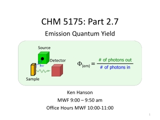
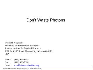
Winfried Wiegraebe Advanced Instrumentation & Physics Stowers Institute for Medical Research 1000 East 50th Street, Kansas City, Missouri 64110 USA Phone: (816) 926-4415 Fax: (816) 926-2088 Email: wiw@stowers-institute.org Don’t Waste Photons
Spectral Imaging: Learn more about your flurochrome Linear Unmixing: Separate overlapping emissions Channel Unmixing: if you can not use a spectral detector Excitation Fingerprinting: Optimize NLO imaging FLIM: Fluorescence Lifetime to distinguish between dyes SHG: Second Harmonic Generation to measure membrane potential FCS: Fluorescence Correlation Spectroscopy – Probe fluctuations to measure diffusion, concentration and interaction (next Technology & Methods Seminar) Don’t Waste Photons
Components of a Laser Scanning Microscope (Pinhole) Detector Laser Beam splitter Scanner Objective Sample
Single Photon Excitation (Confocal Microscope) 1-Photon Focal Region Objective • Out of Focus excitation • Pinhole provides optical sectioning Pinhole Detector
Multiphoton Excitation (Nonlinear Excitation, NLO) 2-Photon 1-Photon Focal Region • 2 photons required for excitation Objective • No out-of-focus excitation • No pinhole required Pinhole • Scattered light is detected Detector
Non-Descanned Detection Pinhole Descanned Detection • No movement of light on detector Scanner Non-Descanned Detection Sample • Light moves on detector • Light moves on sample
Absorption and Emission Spectra Lichtman, J. W. and J.-A. Conchello (2005). "Fluorescence microscopy." Nature Methods 2(12): 910-919.
Spectral Detection • 32 channel PMT • Special grating as dispersive medium • Spectral resolution: 10.7 nm
Photo Conversion of KikGR 561nm: 1.1% 488nm: 3.1% (15x 405nm: 2%) Channel UnmixingDanny.mdb/102705-spec-t channel unmix
Fly Larva expressing ELAV-eGFP • Plan-Apochromat 20x/0.75 • 920nm, 75% • 32 channel META detector FlyLarva012706.mdb/Flylarvalambda@920unmixedfilter.lsm
= a × + b × Linear Unmixing GFP YFP
Linear Unmixing: Fly Larva expressing ELAV-eGFP • Linear unmixing: • eGFP • Autofluorescence • Plan-Apochromat 20x/0.75 • 920nm, 75% • 32 channel META detector • 3x3 lowpass FlyLarva012706.mdb/Flylarvalambda@920unmixedfilter.lsm
Non-Descanned Detector: Fly Antenna expressing ELAV-eGFP • NDD + Transmission: • DIC • NDD2: BP 575-640 • NDD3: BP 500-550 • Plan-Neofluoar 40x/1.3 Oil • 920nm, 25% • 3x3 Lowpass Fly01306.mdb/2ndAntenna@9202channelHBOoff0.lsm
Channel Unmixing: Fly Antenna expressing ELAV-eGFP • Channel Unmixing: • eGFP • Autofluorescence • Plan-Neofluoar 40x/1.3 Oil • 920nm, 25% • NDD2: BP 575-640 • NDD3: BP 500-550 • 3x3 Lowpass Fly01306.mdb/2ndAntenna@9202channelHBOoff2.lsm
Channel Unmixing: Fly Brain expressing ELAV-eGFP • Channel Unmixing: • eGFP • Autofluorescence • Transmitted • Plan-Apochromat 10x/0.45 • 920nm, 32% • NDD2: BP 575-640 • NDD3: BP 500-550
Excitation Fingerprinting: Fly Larva expressing ELAV-eGFP • Plan-Apochromat 20x/0.75 • Ch2 BP 480-520IR • 850 – 950 nm • Excitation fingerprint FlyLarva012706.mdb/Flylarvaexitationseriesfilter.lsm
Timescales in Fluorescence Lichtman, J. W. and J.-A. Conchello (2005). "Fluorescence microscopy." Nature Methods2(12): 910-919.
FLIM: Fluorescence Life Time Imaging Pulsed Laser Detector Dye Molecule Photon Photon Electron Δ t Number detected photons Time delay between laser pulse and detected photon
FLIM: Fluorescence Life Time Imaging Pulsed Laser Detector Dye Molecule Photon Photon Electron Δ t Number detected photons Time delay between laser pulse and detected photon
FLIM: Fluorescence Life Time Imaging Pulsed Laser Detector Dye Molecule Photon Photon Electron Δ t Number detected photons Time delay between laser pulse and detected photon
FLIM: Fluorescence Life Time Imaging Pulsed Laser Detector Dye Molecule Photon Photon Electron Δ t Number detected photons Time delay between laser pulse and detected photon
2PE Fluorescence vs. Second Harmonic Generation (SHG) mouse ovary http://www.drbio.cornell.edu/Infrastructure/NonlinearMicroscopies_WWW/SHG.htm
Fish Scale (Second Harmonic Generation) Sample by Peter Kestler
FLIM: SHG vs. Fluorescence • Second Harmonic Generation in Fish Scale • Fluorescence Lifetime of Fluorescine
Excitation: 850nm → SHG: 425nm • C-Apochromat 40x/1.2 W • 850nm, 5% • 32 channel META • Fish scale SHG101805.mdb/FishscaleMETA800nm.lsm
SHG to Measure Membrane Potential Figure 3 Daniel A. Dombeck et al.: J Neurophysiol (August 10, 2005).
Available: Spectral Imaging: Zeiss LSM 510 META, Leica SP Linear Unmixing: Zeiss LSM 510 META Channel Unmixing: all multi-channel systems Excitation Fingerprinting: Zeiss LSM 510 NLO Special applications: FLIM: Zeiss LSM 510 NLO + B&H FLIM FCS: Zeiss LSM 510 + ConfoCor 3 Future: SHG: Zeiss LSM 510 NLO + special detection optics Don’t Waste Photons
Thanks! • Adv. Instrumentation & Physics • Joseph Huff • Whitney Bartlow • Imaging Facility • Paul Kulesa • Joel Schwartz • Cameron Cooper • Sarah Smith • Danny Stark • Jessica Teddy • Kausik Si • Jeffrey Cotitta, Lisa Sandell, Paul Trainor