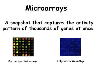
Microarrays: Tools for Proteomics
210 likes | 245 Vues
Explore the world of microarrays and their role in proteomics, focusing on the technology behind these chips and their applications in analyzing diseases like prostate cancer. Learn about the functions of proteins and the methods used in proteomic research. Discover how microarrays are made, their types, and examples of their use in drug discovery, gene expression, and disease identification. Dive into a detailed prostate cancer case study using microarrays to analyze serum immunoreactivity. Uncover the advantages of using microarrays for high-throughput protein analysis.

Microarrays: Tools for Proteomics
E N D
Presentation Transcript
Microarrays: Tools for Proteomics Presented by: Joseph D Gong Chem 395 4/12/05
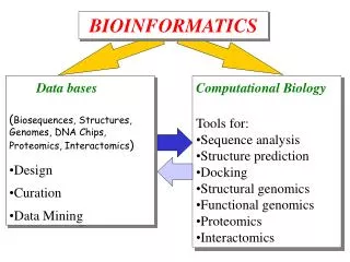
Overview • Introduction • What are chips/micro-arrays? • How are they made? • Applications • Example of an application (prostate cancer) • Conclusions • Questions
Proteins: Peptides Proteins come from subunits called amino acids Amino acids can form short chains called peptides Macromolecules of peptides form to make proteins http://www.catonlimo.com
Functions of Proteins Why are proteins important? • They are important molecules in biological systems • Some major roles they play: • Building blocks of bio-structures (ex. Cell Membranes) • Transport/ Storage of nutrients (ex. Hemoglobin) • Enzymes (metabolism) • Antibodies formation
Proteomics What is proteomics? • The Study of all proteins that are expressed at a certain time insidecells, tissues, organs, or organisms. • Protein Profiling includes: • Abundance, interactions, activity, modifications GOAL: To do this fast and accurately
Proteomic methods • Two Dimensional Gel Electrophoresis (2DE) • Separates numerous amounts of proteins by Size and Charge at varying pH (Old technology) • LC/MS • Protein identification, serum analysis • Microarrays
Microarray What is it? • A slide or chip that contains numerous amounts of biomolecules in fixed amount of space Picture from: www.acefesa.es/microarray/asper/asper.htm
Microarrays How are they made? • Non-contact printing • Piezoelectric • Syringe Solenoid • Contact Printing www.genomicsolutions.com
Contact vs. Non-contact Rose D. A Systems Approach to Fabricating and Analyzing DNA Microarrays. In Microarray Biochip Technology; Schena, M. Ed.; Eaton: Natick, MA, 2000.
Microarray • 3 main types of Microarrays • DNA - genomics • Cell • Protein • Antibody arrays – detects proteins • Protein arrays – detects interactions of proteins or with small molecules
Microarray So what can they be used for? Some examples are: • Drug Discovery/ Toxicology • Gene Expression • Pathogen analysis • Identifying diseases • Cancers • Allergies • Etc.
Prostate Cancer Facts: • Prostate cancer is one of the most common cancers in Men • In 2005 it is estimated that 230,000 new cases of prostate cancer will be diagnosed in the US • Prostate cancer is the second largest cause of cancer death in the US (lung cancer is the first) • 1 out of every 6 men will be diagnosed with prostate cancer in his life time • 1 out of every 33 men will die of this disease Adopted from: Amercian Cancer Society
Example • 1760 fractions of LNCaP cells were collected using 2D liquid Chromatography (Rotofor/RP-HPLC) • Fractions along with several control proteins were spotted in microarrays on nitrocelluslose-coated microscope slides • Sera from 25 men with and 25 men without prostate cancer were incubated individually each on separate microarrays • Immunoglobulins from the sera that bound to spotted fractions were detected after incubating the microarrays w/ biotinylated anti-human Ig and phycoerythrin-streptavidin conjugates (flurophores) and then scanned for flourescence. • This was performed on all 50 microarrays Bouwman, K. et al. Microarrays of tumor cell derived Proteins uncover a distinct pattern of prostate cancer serum immunoreactivity. Proteomics 2003, 3, 2200-2270
Example: Continued • Shows multiple spots with flourescence above the background • An average of 149 (including 15 control proteins) fractions per array showed measurable signal above background Bouwman, K. et al. Microarrays of tumor cell derived Proteins uncover a distinct pattern of prostate cancer serum immunoreactivity. Proteomics 2003, 3, 2200-2270
Example: Continued • Data from all 50 microarrays were grouped and clustered by similarity in intensity patterns • Left 25 columns represent prostate cancer sera from one microarray, Right 25 non-cancer sera • Color: • Red- High intensity • Green- Low intensity • Gray- no data Bouwman, K. et al. Microarrays of tumor cell derived Proteins uncover a distinct pattern of prostate cancer serum immunoreactivity. Proteomics 2003, 3, 2200-2270
Example: Continued • 40 fractions had the most reactivity • 38 fractions had higher reactivity in the prostate cancer sera and only 2 fractions were higher in non. • Many fractions contained the same proteins due to consecutive fraction collection • Further analysis with MS would clarify the number of immunogenic proteins Bouwman, K. et al. Microarrays of tumor cell derived Proteins uncover a distinct pattern of prostate cancer serum immunoreactivity. Proteomics 2003, 3, 2200-2270
Example: Continued • Illustrates the higher level of binding from the prostate cancer sera compared to control sera • Intensities can be quantified Bouwman, K. et al. Microarrays of tumor cell derived Proteins uncover a distinct pattern of prostate cancer serum immunoreactivity. Proteomics 2003, 3, 2200-2270
Advantages High Throughput (Rapid method sample analysis and can handle large samples) Can be used to address protein identification, quantification, and activity studies Can facilitate the discovery of new biomarkers and new drug targets Disadvantages Protein arrays are more complex than DNA/other arrays due to complexity in protein structure Not always direct correlation between protein activity and abundance Stability of proteins to array surface Detection of interacting proteins still weak Advantages/Disadvantages of Microarrays
Conclusions • In example: Strong fluorescence signal from many fractions has sufficient selectivity to detect the binding of specific antibodies proving the usefulness of microarrays • Combined with MS technology, this will further aid characterization and validation of the proteomic method • Although Protein Microarrays have their use in high throughput screening they need to demonstrate better precision, accuracy, and reliability before used in clinical diagnostics
References • http://www.catonlimo.com • www.acefesa.es/microarray/asper/asper.htm • www.genomicsolutions.com • Rose D. A Systems Approach to Fabricating and Analyzing DNA Microarrays. In Microarray Biochip Technology; Schena, M. Ed.; Eaton: Natick, MA, 2000. • Bouwman K. et al. Microarrays of tumor cell derived Proteins uncover a distinct pattern of prostate cancer serum immunoreactivity; Proteomics 2003, 3, 2200-2270 • Talapatra A. Protein Microarrays: Challenges and Promises; Pharmacogenomics 2002, 3(4), 1-10 • Poetz O. et al. Protein Microarrays: Catching the Proteome; Mechanisms of Aging and Development 126 (2005) 161-170
Questions ?’s