Genetic Analysis of Cell Death Pathways in Maize Leaves: Insights from the Camouflage1 Mutant
This study by Huang and Braun, published in the American Journal of Botany, investigates the cellular mechanisms underlying cell death in maize (Zea mays) leaves, specifically focusing on the camouflage1 (cf1) mutant. Utilizing DAB staining methods in both bright-field and UV light, the research highlights distinct pathways involved in cell death, emphasized by contrasting results between wild-type and cf1 yellow tissues. The findings contribute to our understanding of the genetic factors governing leaf senescence and stress responses in maize.

Genetic Analysis of Cell Death Pathways in Maize Leaves: Insights from the Camouflage1 Mutant
E N D
Presentation Transcript
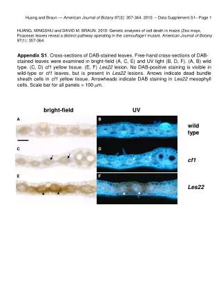
Huang and Braun — American Journal of Botany 97(2): 357-364. 2010. – Data Supplement S1– Page 1 HUANG, MINGSHU and DAVID M. BRAUN. 2010. Genetic analyses of cell death in maize (Zea mays, Poaceae) leaves reveal a distinct pathway operating in the camouflage1 mutant. American Journal of Botany 97(1): 357-364. Appendix S1. Cross-sections of DAB-stained leaves. Free-hand cross-sections of DAB- stained leaves were examined in bright-field (A, C, E) and UV light (B, D, F). (A, B) wild type. (C, D) cf1 yellow tissue. (E, F) Les22 lesion. No DAB-positive staining is visible in wild-type or cf1 leaves, but is present in Les22 lesions. Arrows indicate dead bundle sheath cells in cf1 yellow tissue. Arrowheads indicate DAB staining in Les22 mesophyll cells. Scale bar for all panels = 100 mm. bright-field UV A B wild type C D cf1 E F Les22