Download
1 / 54
540 likes | 744 Vues
Patofyziology of lipids, proteins, aminoacids and purins. Pathophysiology of obesity. Prof. Jan Hanacek. LIPIDS – important part of structural and functional systems of the human body – important part of nutrition

E N D
Patofyziology of lipids, proteins, aminoacids and purins Pathophysiology of obesity Prof. Jan Hanacek
LIPIDS – important part of structural and functional systems of the human body – important part of nutrition – the most important sorce of energy – are dynamicaly changed structures – a lot of them are essential for metabolic processes, others are dangerous They are divided into three main groups: – triglycerides energy production – phospholipids creaqtion of structural and – cholesterol functional molecules, transport of signals in the cells
Functions of some lipids Sphingolipids – play an essential role in maintaining of normal skin function • ceramides – are required for the normal permeability of skin – they create permeability barrier which prevents transcutaneous water loss and penetration of harmful drugs from the environment(antigens) Example of disorder – patients suffering from atopic dermatitis have significantly decreased amount of ceramides in the skin permeability of skin for antigens
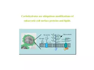
Lipid's caveole and rafts – structural units of biologic membranes – membrane microdomains enriched in sphingolipids and cholesterol – part of plasma membrane signaling machinery – they swimm in more fluid phase of membranes created by glycerophospholipids Functions of lipid rafts They play key role in – transcytosis and endocytosis – signal transmission – internalisation of toxins, viruses and bacterias – cell calcium homeostasis
Nutritional lipids – with saturated fatty acids (bed) – with polyunsaturated fatty acids(good) Transport of lipids in the body – in form of lipoproteins (LPs)(95%) – in form of free fatty acids (FFA) Composition of LPs – TAGs,Cholesterol, Phospholipids, Proteins Classification of LPs according their density – very low density (VLDL) – intermediate density (IDL) – low density (LDL) – high density (HDL)
I. Disturbancies of lipid metabolism Essential types of disturbancies 1. Hyperlipoproteinemias 2. Hypolipoproteinemias 3. Dyslipidemias
Essential terms a)Lipoproteins – spheric particles transporting non-polar lipids (TAGs, cholesterol esters)by blood Composition and properties – inside of sphere - non-polar lipids – surfice of sphere -polar molecules (phospholipids, non- esterified cholesterol- are important for transport of particles in plasma -apo-LPs - are important for LPs metabolism Different types of LPs differs by their density, by volume of transporting lipids, by size, by amount and kind of apo, by location of their creation, by their metabolism
Characteristics of main types of LPs • Chylomicrons(CM) – the lowest density, the largest size • VLDL – smaller and more dense than CM – they transport endogenous TAGs synthetised in liver, mainly • IDL – particles with propertiesbetween VLDL and LDL • LDL – containe cholesterol esters, mainly • HDL – the smallest size and the highest density – they are able to transport cholesterol from peripheral tissues to liver (reversal transport of cholesterol) Lipoprotein (a) – important risk factor for development of atherosclerosis
b)Enzyms important in lipids metabolism • Lipoprotein lipase (LPL) –releses FFAs from TAGs and from VLDL – it is present in endothelial cells – it is activated by Apo C II (it is present in CM and VLDL) • Liver lipase (LL ) – it hydrolyses TAGs in the liver – it is activatedby interaction with Apo E • Lecithin-cholesterol-acyl transferase (LCAT) • Cholesterol ester-transfer-protein (CETP)
• LDL receptor – it takes up LDL (IDL), it is localised at cells in different types of tissues, predominantly at hepatocytes In predispose patients – intake of cholesterol down regulation of LDLr in liver uptake of LDL from blood • HDL receptor – it takes off HDL from blood, it is localised predominantly in cells created steroids – tropic hormons from anterior hypophysis stimulates creationof HDL
Scavenger receptors (SR) – uptake the LDL which were not bind by LDLr – uptake of oxidized LDL particles – they are present in macrophages, in smooth muscle cells in vessel wall atherogenesis
Hyperlipoproteinemias • Definitions: Pathologic process manifested by • concentration of one or more types of LPs • in the blood • Hyperlipidemias –concentration of lipids • in the blood (usually TAGs+Ch) • Dyslipoproteinemias – disorder in lipid spectrum • in blood, usually with • increased concentration of • cholesterol
Hypercholesterolemias - concentration of Ch in blood • It is dengerous situation for the organism: • – 75% of blood Ch is LDL cholesterol • – LDL cholesterol is atherogenic • – atherogenity of LDL cholesterol increases with the • degree of its oxidation and glycation • – oxidized and glycated LDL are taking off by SR on • the surfice of macrophages and smooth muscle cells • development of foam cells • b) Hypertriacylglycerolemias • c) Combination of a) and b)
Classification of hyperlipoproteinemias (according Necas et al., 2000)
Main types of hyperlipoproteinemias (HLP) • Primary • 1.Familial combined HLP • – it is the most frequent genetic HLP • – it manifests most likely in phenotypes 2a, 2b, 5 • – it acompanies metabolic X syndrome • – it is the strong risk factor for development of • atherosclerosis and ischemic heart disease • Mechanisms involved in development HLP • • genetic predisposition • • acquired (due to environmental factors) secretion of VLDL by liver
2. Familial hypercholesterolemia (FHC) – it manifests predominantly by phenotype 2a – it leads to significant acceleration of atherosclerosis development – myocardial infarction in 4th decade of life – xantomatosis of tendons and arcus lipoides corneae Mechanisms involved in FHC development – mutation of LDL receptor decreased uptake of LDL concentration of LDL in blood
3. Polygenic hypercholesterolemia- the most frequent hypercholesterolemia (type 2a) – there are not xantoms – in 1st line relatives in family is lower frequency of hypercholesterolémia than in 2nd types of HC Mechanisms of development –genetic predisposition – changes of resorbtion and endogenous synthesis of cholesterol, changes in metabolism of LDL, other changes – environmental factors – alcohol, DM, intake of carbohydrates and lipids
4. Familial dyslipoproteinemia – there is significant xantomatosis and acceleration of atherosclerosis – it manifests in form type 3 HLP Mechanism of development – polygenic disturbance 5. Familial hyper TAG– quite frequent disorder – concentration of Ch in blood must not be increased – it manifests in form type 4 HLP Mechanism of development – inherited disorder
6. Familial defect oflipoprotein lipase and Apo C II – rather rare genetic disorder – in homozygotes – accumulation of TAGs in tissues, xantoms, hepatosplenomegaly, high risk of acute pancreatitis – it manifests by phenotype 1 (when defect of LPL) or by phenotype 5 (when defect of Apo C II ) 7. Familial hyperalfalipoprteinemia – concentration of HDL in blood risk of atherosclerosis development Mechanisms of development – genetic disorder – low doses of alcohol – estrogens
B. Secondary – are induced by other kind of disease The most frequent diseases accompanied by HLP – diabetes mellitus – nephrotic syndroma – chronic renal failure – hypothyreosis – primary biliary cirhosis – chronic alcoholism – some drugs, e.g. contraceptives
The role of lipidrafts in pathogenesis Disorders of structure and function of lipid rafts is involved in pathogenesis of: – virus infections – e.g. HIV – Alzheimer disease, Parkinson disease – prionoses, e.g. Creutzfeldt- Jakob's disease – immunity disorders, e.g. allergy – tumors – atherosclerosis – systemic hypertension - others
II. Disorders of protein and aminoacids metabolism • Disorders of nitrogen balance • a) positive nitrogen balance • – growth, convalescens, gravidity, sportsmen... • b) negative nitrogen balance • – catabolic processes, e.g. chronic diseases as are • CHOLD, cancer, fever, nutritional disorders • 2. Disorders in blood protein spectrum • a) production of monoclonal immunoglobulins, • – e.g. Waldenstrom's macroglobulinemia blood • viscosity • Mechanism: production of IgM
– e.g. multiple myeloma blood viscosity Mechanism: production of IgA b) production of cryoglobulins disorders of microcirculation Mechanism: – cryoglobulins precipitation when temperature of blood will decrease (peripheral blood) c) hyperfibrinogenemia, cryofibrinogenemia – disorders in hemocoagulation d) hypoalbuminemia– due to liver and renal diseases
3. Disorders of aminoacids metabolism a) Phenylketonuria – Phenylalanin – essential AA tyrosine creation Mechanism: - mutation of gene for phenylalanin hydroxylase Consequences: – phenylalanin accumulation onset of abnormal metabolits creation, e.g. phenyl pyruvate, phenylacetate – damage of nerve system – hypopigmentation ( due to ihibitory influence of phenylalanin on melanin creation
b) Albinism – decreased amount or absence of melatonin in skin, hairs and eye Mechanism: – defect of tyrosinase enzyme Consequences: – oculocutaneous albinism – increased sensitivity of skin to UV radiation – photophobia and vision disorders
c) Alkaptouria (ochronosis) – disorders in metabolism of phenylalanin – homogentisic acid is created by metabolisation of phenylalanin – there is defect of oxidation of homogentisic acid Consequences: – accumulation of brown-red pigment in connective tissues (ochronosis) – damage of joints cartilage arthrosis – damage of heart valves – valvular heart disease – excretion of pigment by urine – brown-red or blue-black color of auricula and sclera
d) Homocysteinuria – accumulation of homocystein in blood due to disturbancies of metabolism of sulphur containing AA Consequences: – concentration of homocystein in blood – damage ofendothelial cells accelerated atherosclerosis – damage of vision
III. Disorders of purin's metabolism – purins: -compounds created nucleic acids (NA) – metabolism of purins uric acid (UA) Hyperurikemia and gout – hyperurikemia – concentration of uric acid in blood – sorces of uric acid – food, NA of the own organism Primary hyperurikemia – the cauce is not clearly known Possible factors involved: – genetic predisposition – limited excretion of UA by kidney – high dose of NA in food – activity of enzymes created AMP, GMP from UA
Secondary hyperurikemia – due to some diseases – renal failure – cytostatic therapy of cancer – Uric acid is well excreted when urine is alkalic – Solubility of UA in synovial fluid decreses with decreasing of its temperature Gout – disease developed due to hyperurikemia and accumulation of urates to the distal joints of foot (microtophi) Pathogenesis – creation of microcrystals of UA in tissues – phagocytosis of microcrystals by LE – cascade of local inflammatory processes
Results: –damage of joints, kidney, vessels – asseptic inflammation of joints and tissues around them deformation of joints – acute urate nephropathy IV. Disorders of porfirin's metabolism – see the Color Atlas of Pathophysiology
Pathophysiology of obesity Prof. J. Hanacek, M.D., Ph. D.
Essential epidemiologic data on obesity • More than 7% world population suffer from obesity • Incidence of overweight and obesity has increased during the last two decades „epidemic of obesity“ • Frequency of obesity is increasing significantly especially in countries with high % of pauperised inhabitants for a prolonged period, when the accesability of food suddenly improved • There is increased incidence of obesity in children • Negative influence of obesity on men health is now convincingly proven
Definition of obesity • We considere as obese the person whose weight is significantly over the upper limit of physiologic range, due to accumulation of fat – in men more then 25%, in wumen more tha 30% of total body weight • Obesity is considered as chronic disease which can result in multiorgan damage manifeted as complications of obesity • Obesity is the result of influence of many pathogenic mechanisms
Physiologic remarks •The preponderance of stored energy consists of fat • Intake of energy and energy expanditure is during longer period of life in balance • Energetic substrates of food are used in the body: – for essential metabolic processes (75%) – for thermogenesis (10-15%) – for exercise (10-15%)
Methods used for diagnosis of obesity • Simple methods: • a) BMI =body weight(kg)/ hight (m2) • Normal BMI: 18,5 – 25 • b) Waist-to-hip ratio • Normal values: 0.7-0.95 men; 0.7-0.85 women • c) Waist circumference: <95 for men; <81 for women • d) Skinfold thickness (on the trunk and extremities) • 2. Sophisticated techniques: • CT, denzitometria, dilutional methods, spectrometry...
Main causes of body weight increse • Muscle mass increase • Body water amount • Body fat mass increse • Expression of overweight degree by BMI: • Overweight:25-30 - (grade 1 • Obesity: 30-35 – (grade 2) • Obesity: 36-40 – (grade 2) • Gross obesity:>40 – (grade 3) • Classification of obesity • Etiopathogenetic- 1. Primary • 2. Secondary
B. Pathologic – anatomy 1. Hypertrophic form 2. Hypertrophic-hyperplastic form C. According fat distribution 1. Android type (apple shaped) – fat localised in trunk and in abdominal cavity – risk of DM, AMI, brain ischemia, other deseases of CVS 2. Gynoid type (pear shaped) – fat localised at gluteal part, at thighs – risk of joints damage, mainly
The main causes and mechanisms involved in obesity development •The essential pathomechanism Caloric intake exceeds for a longer time the energy expanditure • The particular mechanisms I. Primary increase of energy intake II. Primary decrease energy expanditure III, Combination of both previous mechanisms
Main groups of causes lading to obesity • Genetic disorders • – about 33% existing forms of obesity is the result • of genes dysfunction • Environmental factors (with some influence of genes) • – socio-economic stress lover level of education, • lover incom, lover cultural • level... • – insufficient physical activity (life style) • – national and regional eating habits • – increased intake of alkoholic beverages (no chronic • alcoholism)
The roles of brain in obesity development •brain controls of caloric intake and energy expanditure • brain structure or/and function disorders can lead to disorders in energy intake and energy expanditure B R A I N Aferent signals – nerv – humoral-metabolic (e.g. insulin, glucose, CCK, specific cytokines) Eferent signals – control of intake – control of expanditure – control of fat mass
• Damage of ventro-medial hypothalamus (VMH) Consequences: – hyperfagia – setpoint for body weight obesity Characteristic features of metabolism: – efficacy of metabolism ( glucose is oxidised, fat is stored) – hyperinsulinemia – increased vagal activity – decreased sympathetic activity • Abnormal function of SNS and PSNS Consequences: activity of SNS in pancreas, heart, fat tissues abnormal thermogenesis
Probably common end-part of pathway in CNS responsible for onseting of obesity • Aberant control of neurons producing NPY (Physiology: -glucose, insulin, leptin... + monoamins in CNS inhibition of NPY production by neurons in n. arcuatus inhibition energy intake) In obese persons: – possible resistance of NPY neurons to aferent metabolic signals NPY production is not inhibited energy intake is not inhibited
Effects of food composition on obesity onset Hypothesis:High concentration of fat in food intake of calories development of obesity Results of research: – satiating efficiency of fat is smaller than carbohydrates and proteins passive overeating – high energy concentration in fat unit of food – fat in meals taste very well facilitation of eating, speed and amont of food intake are increased – later development of satiating signal during eating fatty meals
Fat paradox: signals of satiety induced by fat intake versus hihg fat hyperfagia Explanation: • If fat come to small intestine strong pre-absorbtivesignalismediated by: – mainly CCK, – also by glukagon, enterostatin, – by products of fat digestion • Signals from energy sorces e.g. satietin, adipsin, leptin... modulation of regulatory circuits in CNS involved in calory intake control
• Intake of fat per os fat will come to small intestine with time lap Result: – less intens and later signals of satiation slowness in decrease of hunger feeling • Fat in mouth intens stimulation of taste receptors facilitation of fat intake nice taste of fat is able overcome the satiation signals coming to CNS • High density of energy in fat intake of large amount of energy till satiation signals are able to inhibit feeling of hunger
Visceral obesity – accumulation of fat in abdominal cavity • Strong relation does exist between visceral obesity and development of metabolic complications Example: 2 groups of obese persons with equal BMI – 1st group: fat localised subcutaneously at trunk – 2nd group: fat localised in abdominal cavity Differences in metabolic parameters: – persons in the 2nd group had values of PGTT and TAG in blood compared with 1st group Increased fat mass in abdominal cavity leads to sensitivity to insulin indipentendly on BMI