Hematology and Immunology
530 likes | 1.18k Vues
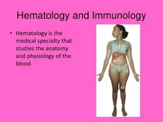
Hematology and Immunology. Hematology is the medical specialty that studies the anatomy and physiology of the blood. Anatomy and Physiology (cont’d). The Blood http://www.youtube.com/watch?NR=1&feature=endscreen&v=CRh_dAzXuoU

Hematology and Immunology
E N D
Presentation Transcript
Hematology and Immunology • Hematology is the medical specialty that studies the anatomy and physiology of the blood.
Anatomy and Physiology (cont’d) The Blood http://www.youtube.com/watch?NR=1&feature=endscreen&v=CRh_dAzXuoU Contains blood cells, blood cell fragments, water, and other substances (proteins, clotting factors, etc.) Transports oxygen, carbon dioxide, nutrients, and waste products Contains cells that also function as part of the immune system
Anatomy and Physiology (cont’d) The Lymphatic System http://www.youtube.com/watch?feature=player_embedded&v=Kh-XdNnTZUo Consists of the lymphatic vessels, lymph fluid, lymph nodes, lymphoid tissues, and lymphoid organs Forms a pathway throughout the body that is separate from that of the cardiovascular system that contains the blood Defends the body against microorganisms and cancerous cells
Anatomy of the Bloodhttp://www.youtube.com/watch?v=R-sKZWqsUpw&feature=related Plasma Clear, straw-colored liquid (about 90% water) that makes up 55% of the blood. The formed elements of the blood (erythrocytes, leukocytes, and platelets) are suspended in the plasma.
Anatomy of the Blood (cont’d) Plasma (cont’d) • Contains substances the body produces itself such as: albumin, bilirubin, hormones, complement proteins, and clotting factors. • Contains creatinine and urea, which are waste products of cellular metabolism.
Anatomy of the Blood (cont’d) Erythrocytes Most numerous of the formed elements in the plasma Red blood cell that is a round, somewhat flattened, red disk Unique because, unlike other body cells, they have no cell nucleus when they are mature
Anatomy of the Blood (cont’d) Erythrocytes (cont’d) Contain hemoglobin, a red, iron-containing molecule that binds to oxygen molecules to form oxyhemoglobin. Hemoglobin carries oxygen from the lungs to every cell in the body, and carries carbon dioxide from the cells back to the lungs. Hematopoiesis, the process by which all blood cells are formed, occurs in the red marrow of long or flat bones.
Hematopoiesis is the process by which all blood cells are formed, it occurs in the red marrow of long or flat bones.
Anatomy of the Blood (cont’d) • Erythrocytes (cont’d) • Very immature cells are known as stem cells. • Erythrocyte stem cells mature to become erythroblasts and then normoblasts.
Anatomy of the Blood (cont’d) Erythrocytes (cont’d) Do not have a nucleus, so they cannot divide or repair themselves. Last 120 days before they begin to deteriorate.
Macrophages, the principal phagocytic (cell-engulfing) components of the immune system, ingest and destroy foreign particles such as bacteria.
Anatomy of the Blood (cont’d) Erythrocytes (cont’d) Iron stripped from heme molecules is stored in the liver and the spleen; the remainder of heme molecules is converted to bilirubin. Bilirubin plays an important role as an antioxidant, protecting body cells from damage by free radicals.
Interesting Facts: Two million red blood cells die every second. There are approximately 100,000 miles of blood vessels in the human body. Seven percent of a humans body weight is made up of blood. Each day 400 gallons of recycled blood are pumped through the kidneys. By donating just one pint of blood, four lives can be saved. Half your body’s red blood cells are replaced every seven days. Vampires prefer type A, warm blood but will settle for other types as long as they are warm. Every two seconds, someone needs blood. There are approximately 1 billion red blood cells in two to three drops of blood. Your body usually replaces the volume of the blood you donate within 24 hours. Restoring red blood cell levels to normal can take up to two months though. If you are healthy, you will be eligible to donate blood up to 330 times in your life.
Anatomy of the Blood (cont’d) • Leukocytes (White blood cells) • include five types of cells (neutrophils, eosinophils, basophils, lymphocytes, and monocytes) • can be identified by the presence or absence of granules in their cytoplasm and the shape of their nucleus
Anatomy of the Blood (cont’d) Leukocytes (cont’d) Leukocytes with large granules in their cytoplasm are categorized as granulocytes, which include neutrophils, eosinophils, and basophils. Leukocytes with few or no granules in their cytoplasm are categorized as granulocytes, which include lymphocytes and monocytes.
Anatomy of the Blood (cont’d) Neutrophils • Most common leukocyte, making up 40 to 60% of leukocytes in blood • Categorized as granulocytes • Nucleus has many segments or lobes, so they are also known as polymorphonucleated leukocytes (PMNs), polys, segs, or segmenters
Anatomy of the Blood (cont’d) Neutrophils (cont’d) Develop in the red marrow Engulf and destroy bacteria (phagocytosis) Live only a few days or even just a few hours if they are actively destroying bacteria
Anatomy of the Blood (cont’d) Eosinophils • Make up just 1 to 4% of leukocytes • Categorized as granulocytes; also known as eos • Nucleus has two lobes • Develop in the red marrow • Engulf and destroy foreign cells (pollen, animal dander, etc.) • Release chemicals that kill parasites
Anatomy of the Blood (cont’d) • Basophils • Least common leukocyte, making up 0.5 to 1% of leukocytes • Categorized as granulocytes; also known as basos • Nucleus has more than one lobe • Develop in the red marrow • Release histamine at the site of tissue injury • Release heparin, an anticoagulant
Anatomy of the Blood (cont’d) • Lymphocytes • Make up 20 to 40% of leukocytes. • Categorized as agranulocytes and are the smallest leukocytes; they are also known as lymphs. • Nucleus is round and nearly fills the cell. • Some lymphocytes live for just a few days, while others live for many years.
Anatomy of the Blood (cont’d) • Lymphocytes (cont’d) • Begin development in red marrow; some become B cells or natural killer cells; others migrate to the thymus to become T cells • Present in the blood and lymph nodes; destroy viruses and produce antibodies
Anatomy of the Blood (cont’d) Monocytes Make up 2 to 4% of leukocytes Categorized as agranulocytes and are the largest leukocytes; also known as monos Have a large amount of cytoplasm, and nucleus is large and kidney bean shaped Develop in the red marrow
Anatomy of the Blood (cont’d) • Monocytes (cont’d) • Are phagocytes that engulf and destroy microorganisms, cancerous cells, dead leukocytes, and cellular debris. • Monocytes in the lymph nodes, intestine, liver, pancreas, thymus, spleen, bone, and skin are known as macrophages.
Anatomy of the Blood (cont’d) • Thrombocytes • Different from other blood cells because they are only cell fragments • Active in the blood-clotting process • Begin in the red marrow as stem cells that then become megakaryoblasts, and then mature into megakaryocytes, a very large cell • Cytoplasm of the megakaryocyte breaks away at the edges to form cell fragments (thrombocytes) that are released into the blood
Anatomy of the Blood (cont’d) • Blood Type • Most important blood types are the ABO and Rh blood groups • ABO blood group contains A, B, AB, and O antigens
Anatomy of the Blood (cont’d) Blood Type (cont’d) Rh blood group has 47 different antigens Rh is positive when antigens are present on erythrocytes Rh is negative when antigens are not present on erythrocytes
Figure 6-10 A unit of blood Shout Pictures/Custom Medical Stock Photo, Inc.
Physiology of Blood Clotting Platelet aggregation―Thrombocytes form clumps to decrease blood loss Coagulation―Blood clot forms Hemostasis―Cessation of bleeding When clotting factors in the plasma are activated to form a blood clot, the fluid portion of plasma that remains is known as serum.
Figure 6-11 Blood clot Susumu Nishinaga/Photo Researchers, Inc.
Anatomy of the Lymphatic System Lymphatic Vessels, Lymph, and Lymph Nodes Lymphatic vessels are similar in structure to blood vessels, but with several important differences. Begin as tiny lymphatic capillaries in the tissues.
Anatomy of the Lymphatic System (cont’d) Lymphatic Vessels, Lymph, and Lymph Nodes (cont’d) End in ducts that empty into large veins in the neck. Tissue fluid enters a lymphatic capillary and becomes lymph, the fluid that circulates through the lymphatic system.
Anatomy of the Lymphatic System Lymphatic capillaries have large openings in their walls that allow microorganisms and cancerous cells to enter. Lymphatic capillaries become larger lymphatic vessels that bring lymph to the lymph nodes. Valves keep the lymph flowing in one direction.
Anatomy of the Lymphatic System (cont’d) Lymphoid Organs (cont’d) Grouped together in chains in areas where there is a high risk of invasion by microorganisms or cancerous cells. Lymphatic vessels end at ducts in the thoracic cavity: right lymphatic duct and thoracic duct. Both lymphatic ducts then empty into large veins in the neck. Physical Barriers
Anatomy of the Lymphatic System (cont’d) Lymphoid Tissues Contain lymphocytes and macrophages that are active in the immune response Tonsils and adenoids in the oral cavity Appendix and Peyer’s patches in the small intestine
Anatomy of the Lymphatic System Lymphoid Organs Thymus is located within the mediastinum and helps lymphoblast's mature into T lymphocytes
Anatomy of the Lymphatic System • Lymphoid Organs • Spleen is located on left side of abdominal cavity and is the largest organ in the lymphatic system • Spleen removes old erythrocytes from the blood • Spleen also acts as storage area for whole blood, which is released into the circulatory system during times of danger or injury