Download
1 / 17
180 likes | 414 Vues
Experimental Design in fMRI. A real example of fMRI block design done well: alternate moving, blank and stationary visual input. Moving. Blank. Stationary. Blank. 40 sec. 40 sec. 40 sec. 40 sec. Experimental Design in fMRI. Voxels in Primary cortex tracked all stimuli.

E N D
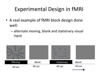
Experimental Design in fMRI • A real example of fMRI block design done well: • alternate moving, blank and stationary visual input Moving Blank Stationary Blank 40 sec 40 sec 40 sec 40 sec
Experimental Design in fMRI • Voxels in Primary cortex tracked all stimuli
Experimental Design in fMRI • Voxels in area MT tracked only the onset of motion
Experimental Design in fMRI • Voxels in area MT tracked only the onset of motion • How did they know to look in area MT?
Structural and Functional Imaging • What you really want is an image, not a squiggly line • Make a map of a statistic (like t-score or z-score) that describes how well each voxel tracked the cognitive task: • Set all “non-significant” voxels to be transparent
Structural and Functional Imaging • What you really want is an image, not a squiggly line • Make a map of a statistic (like t-score or z-score) that describes how well each voxel tracked the cognitive task: • Set all “non-significant” voxels to be transparent
PET: another way to measure blood Oxygenation • Positron Emission Tomography (PET) • Injects a radioisotope of oxygen • PET scanner detects the concentration of this isotope as it decays
Advantages of fMRI • Advantages of MRI: • Most hospitals have MRI scanners that can be used for fMRI (PET is rare) • Better spatial resolution in fMRI than PET • Structural MRI is usually needed anyway • No radioactivity in MRI • Better temporal resolution in MRI
Advantages of PET • Advantages of PET: • Quiet • A number of different molecules can be labeled and imaged in the body
Limitations of fMRI • All techniques have constraints and limitations • A good scientist is careful to interpret data within those constraints
Limitations of fMRI • Limitations of MRI and PET: • BOLD signal change does not necessarily mean a region was specifically engaged in a cognitive operation • Poor temporal resolution - depends on slow changes in blood flow • expensive
Neurons are Electrical • Remember that Neurons have electrically charged membranes • they also rapidly discharge and recharge those membranes (graded potentials and action potentials) • Review relevant textbook sections if this isn’t familiar to you
Neurons are Electrical • Importantly, we think the electrical signals are fundamental to brain function, so it makes sense that we should try to directly measure these signals • but how?
Subdural Grid • Intracranial electrodes typically cannot be used in human studies
Subdural Grid • Intracranial electrodes typically cannot be used in human studies • It is possible to record from the cortical surface Subdural grid on surface of Human cortex
Electroencephalography and the Event-Related Potential • Could you measure these electric fields without inserting electrodes through the skull?