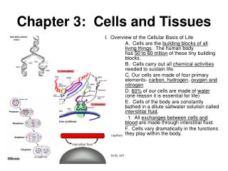

The Cell: Units of Life and Cell Membrane
E N D
Presentation Transcript
A. Cells are the units of life • All organisms consist of cells • Cells are the smallest unit of life that can function independently • 1660 – Robert Hooke first person to see outlines of cells (in cork) • 1673 – Antony van Leeuwenhoek improved lenses and drew observations • 1830’s – Robert Brown identified the nucleus
Cell theory (est. ~ 1839) • 2 Main tenets (ideas) • All organisms are made of one or more cells • The Cell is the fundamental unit of life • Ideas developed by Schleiden and Schwann • 3rd Main tenet • All cells come from preexisting cells • Contradicts the idea of spontaneous generation • This idea was added in 1855 by Virchow
Microscopes are used to study cells • Light microscopes • Compound light microscope - glass lenses focus visible light, 0.2 µm resolution • Confocal microscope – enhanced resolution using white or laser light
Electron microscopes – greater magnification and better resolution but specimen must be dead • Transmission Electron Microscope (TEM) – uses beam of electrons focused by magnetic field • Scanning Electron Microscope (SEM) – scans beam of electrons over metal coated specimen • Scanning probe microscope – probe moves over surface giving exquisite detail
Features common to all cells • Genetic information, DNA • Proteins carry out cell’s work • RNA participates in producing proteins • Ribosomes manufacture proteins • Cytoplasm • Cell membrane • Complex cells also have organelles – compartments for specialized functions
Surface area to volume • All cells are small • Require large surface area • Surface area limitation on size of cell • May be avoided through • Flattened shape • Fingerlike extensions • Specialized organelles to improve efficiency (explains animal and plant cells being larger than bacterial cells) • Vacuoles in plant cells, etc.
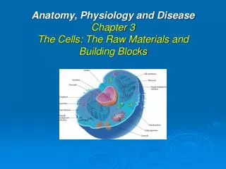
B. Cell membrane (separating the cell from the external environment and So much more!) • Composition of the cell membrane: • Made of lipids and proteins • Phospholipid • Glycerol and a phosphate group form head, 2 fatty acids form the tails • Head is hydrophilic, tails are hydrophobic • Together, phospholipids spontaneously form phospholipid bilayer, like a defense move for the tails! • Fluid mosaic – proteins and phospholipids free to move laterally within the bilayer
Proteins in the cell membrane: • Transport proteins • Adhesion proteins • Enzymes • Receptor proteins • Recognition proteins
Functions of the Cell Membrane: • Signal transduction • A cell receives an external “message” and converts it into an internal signal • Receptor proteins bind to stimulus molecule, first messenger • Triggers chemical reaction whose product is second messenger • Second messenger provokes cell’s response – activating particular genes or enzymes
C. Different cell types • Prokaryote – simplest and most ancient forms of life whose cells lack organelles • Eukaryotes – cells contain organelles • 3 domains of life • Bacteria – prokaryote • Archaea – prokaryote • Eukarya – eukaryote
C. Different cell types • Domain Bacteria • Lack membrane bound nuclei • 1 circular DNA molecule found in nucleoid • Rigid cell wall in most – provides protection and shape • Some have capsule and flagella • Domain Archaea • Resemble bacteria superficially only • Phospholipids, cell walls, and flagella unique • Some are “extremophiles” • Burning Question: What IS the smallest certifiable living organism? See p. 59!
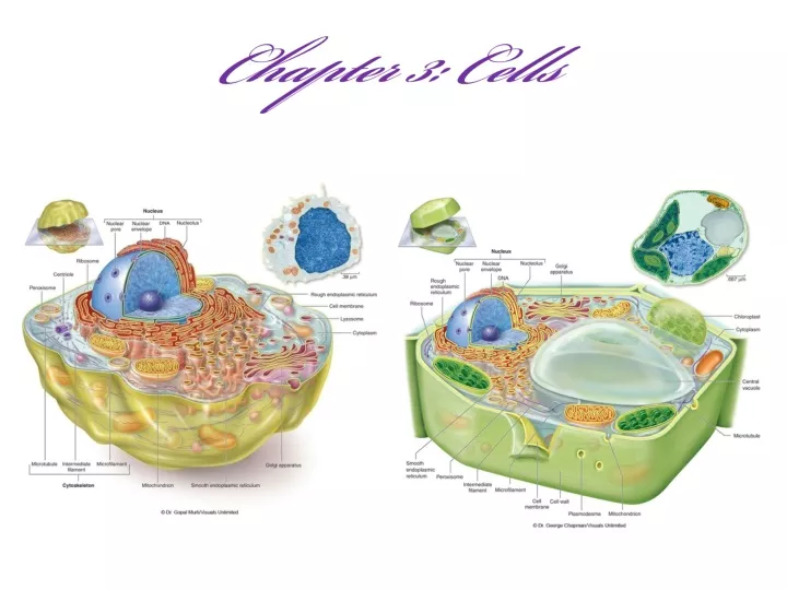
C. Different cell types • Domain Eukarya • Huge diversity • Larger than prokaryotes • Internal membranes create internal compartments called organelles that are specialized for specific tasks in the cell • Endosymbiosis theory – maybe ancient organism engulfed another organism and rather than eating it, it stayed as a partner (supported by mitochondria and chloroplasts) • 2 Major groups of eukaryotic cells based on structure and functions: animal & plant cells
D. Eukaryotic organelles • Organelles carry out coordinated interactions to meet the needs of an organism: Ex: Milk
D. Eukaryotic organelles • Nucleus • Contains DNA – information specifying “recipe” for every protein a cell can make • Nuclear pores are holes in the nuclear envelope surrounding the nucleus, lets RNA and other substances through • Nucleolus - dark core in middle of nucleus; assembles ribosomes • Cytoplasm • Watery-jelloish soup of dissolved substances, organelles and cytoskeleton
D. Eukaryotic organelles • Ribsomes • Site of protein synthesis • Found lose in cytoplasm or attached to ER • Endoplasmic Reticulum • Originates at nuclear membrane and winds throughout the cell • 2 types: • Rough ER – studded with ribosomes making proteins destined for secretion (like milk) • Proteins folded and modified • Smooth ER – synthesizes lipids, detoxifies drugs and poisons • Lipids and proteins made by ER exit in vesicles
D. Eukaryotic organelles • Golgi apparatus • Processing center for vesicle contents • Proteins complete intricate folding and become functional • Some proteins will become membrane surface proteins • Others packaged for secretion from the cell
D. Eukaryotic organelles • Lysosomes • Contain enzymes that lyse substrates • Specific pH inside lysosome prevents enzymes from damaging cell • Vacuoles • Found in plants • Enzymes degrade and recycle materials • Important in growth and maintaining rigidity • Peroxisomes • Dispose of toxic substances • Some reactions produce hydrogen peroxide (H2O2) • Enzyme produces harmless water molecules
D. Eukaryotic organelles • Chloroplasts • Found in plant cells • Site of photosynthesis • Uses energy from sunlight to produce glucose • Occurs specifically in thylakoids • Endosymbiosis theory support – has its own DNA
D. Eukaryotic organelles • Mitochondria • Cellular respiration extracts energy from food • Cristae – internal folds; contain enzymes for cellular respiration • Also contains its own DNA • Always inherited from mother in humans http://www.pbs.org/wgbh/nova/neanderthals/mtdna.html
3 major components distinguished by protein type, diameter, and aggregation Microtubules Microfilaments Intermediate filaments Cytoskeleton D. Eukaryotic organelles
Microtubule Made of Tubulin protein Forms very small hollow tubes Can change length of tube by adding or removing tubulin molecules “Trackway” within cell for many cellular movements Cilia – short, many Flagella – long, few D. Eukaryotic organelles
D. Eukaryotic organelles • Microfilaments • Actin • Long, very thin rods • Machinery to move • Intermediate filaments • (10 nm) diameter is intermediate • Made of different proteins in different specialized cell types • Internal scaffold for cell
Cell walls Surround cell membrane of nearly all bacteria, archaea, fungi, algae, and plants Not just a barrier Built of different components Plasmodesmata connect adjacent cells (like little bridges between cells) E. Cells adhere and communicate
Animal cell junctions Animal cells lack cell walls Secrete complex extracellular matrix Intercellular junctions Tight junctions form impermeable barriers Anchoring or adhering junctions connect cells by linking intermediate filaments Gap junctions link cytoplasm of adjacent cells http://www.cellsalive.com/cells/3dcell.htm http://learn.genetics.utah.edu/content/begin/cells/insideacell/ http://learn.genetics.utah.edu/content/begin/cells/scale/ http://www.wisc-online.com/objects/index_tj.asp?objID=ap11604