Download

1 / 25
250 likes | 458 Vues
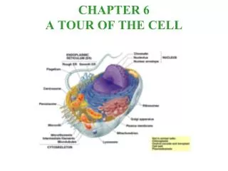
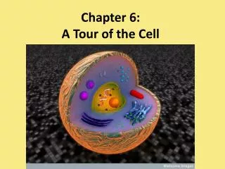
Chapter 7 A tour of the Cell. What do all cells have in common?. Prokaryotic Cells. Eukaryotic Cells. Eu = true; Karyon = nucleus Cells w/ nucleus Linear DNA contained in nucleus 10 – 100 micrometers in size With membrane-organelles (endomembrane system) Cell wall only in plants

E N D
What do all cells have in common? Prokaryotic Cells Eukaryotic Cells • Eu = true; Karyon = nucleus • Cells w/ nucleus • Linear DNA contained in nucleus • 10 – 100 micrometers in size • With membrane-organelles (endomembrane system) • Cell wall only in plants • Ex. Plants and animal cells • Pro = before; Karyon = nucleus • Cells w/o nucleus • Circular DNA free in “nucleoid” region • 1 – 10 micrometers in size (size of mitochondria) • No membrane organelles • Cell wall • Ex. bacteria • Phospholipid plasma membrane • Cytoplasm • Ribosomes • Genetic Material (DNA)
Animal Cells Plant Cells
Nucleus • Contain DNA of eukaryotic cells • Porous phospholipid membrane • Inner membrane lined with intermediate filaments of the cytoplasm • ER often is an extension of the nuclear membrane
Condensation of Eukaryotic Chromosomes Nucleosome = DNA coils around histone proteins Chromatin = supercoiled nucleosomes Looped Domains = supercoiled chromatin Chromosome = supercoiled looped domains
Ribosomes • Assembles AA into polypeptide chain, which eventually folds into functional protein • Made of rRNA and protein • 2 subunits: large and small
Nucleolus • Located inside nucleus • makes ribosomal subunits by combining rRNA and proteins imported from cytoplasm • subunits leaves nuclear pore and assembles into a ribosome in the cytoplasm
What is the endomembrane system? • System of membrane-bound organelles in euk. cells that work cooperatively together to create secretory proteins, membrane-bound proteins, or plasma membrane proteins • Nucleus • ER • Golgi • Transport Vesicles • Lysosomes • Peroxisomes • Vacuoles • Plasma Membrane
Rough Endoplasmic Reticulum RER w/ bound ribosomes Space w/in ER = cisternae space Fcn: to fold and modify secretory proteins (glycoproteins) within cisternae space - attaches CHO called oligosaccharides to growing and folding polypeptide chain (2o 3o) - vesicles bud off from RER and delivers glycoprotein to Golgi
Golgi Apparatus Accepts vesicles from RER (cis side) Adds and removes monomers to oligosach. of glycoproteins Adds “ID” tags (like phosphate groups) and uses these to “sort” proteins into different vesicles Dispatches vesicles w/glyco-proteins for shipping (trans side)
3 destinations for proteins within Golgi vesicles • Secreted from cell • Remains within vesicles vacuole, lysosome, peroxisome • Protein becomes part of plasma membrane
Lysosomes • Membrane-bound sac of digestive enzymes • Acidic env’t maintained by pumping H+ions from cytoplasm • Digests food, worn out cell parts, programmed cell death (webbing b/t fingers, tadpole tails)
Peroxisome • Breaks down toxic substances in liver • Breaks down fatty acids into CHO for use in CR • In breakdown process, oxygen and hydrogen combine to create H2O2 • Peroxide = metabolic waste
Smooth ER • ER w/o ribosomes • Makes lipids, oils, steroids • Helps break down CHO • Detoxifies drugs by adding –OH groups water soluble toxins flushed from body
Contractile vacuole - pumps excess water out for freshwater organisms Central vacuole - Stores water, organic compounds, ions, and helps increase turgor pressure in plant cells Vacuoles
Mitochondria CR site Generates ATP (usable E) from glucose Chloroplasts PS site Generates glucose (stored E) from inorganic compounds and light Mitochondria and Chloroplasts
Cytoskeleton Network of fibers in the cytoplasm that a) maintains cell shape/mechanical support b) anchors organelles c) helps w/ cell motility 3 components 1) microtubules 2) microfilaments 3) intermediate filaments
Microtubules Structure: Hollow tube made up of α and β tubulin polypeptide 25 nm diameter Compression Resistent supports cell shape Forms spindle fibers for separation of chromosomes, makes up centrioles, and cilia/flagella
9 sets of 3 arrangement (ring formation) Ex. Centrioles, spindle fibers, basal body of cilia and flagella 9 + 2 arrangement (9 doublets surrounding a pair in the center) Ex. Cilia and Flagella Microtubule
Radial Spokes and Dynein Arms of Microtubule • Dynein arms “walk” along the microtubules to bend and move flagella, using ATP energy
Microfilaments AKA: actin fibers Structure: twisted double chain of actin protein that forms a solid rod 7 nm diameter Tension resistent (protects against “pulling” forces) Makes up microvilli core, contracts muscles, causes cytoplasmic streaming and pseudopod extensions in cells
Intermediate Filaments • In btwn microtubules and microfilaments in size (10 nm) • Fixes positions of organelles • Organelles w/motor proteins can move by “walking” along intermediate filaments (as if along a track) • Helps to maintain cell shape
Cell Wall and Plasmodesmata • In plants, bacteria, protists, fungi • Maintains shape, prevents excessive water uptake • Cell walls are non-continuous: plasmodesmata connects cytoplasm btwn 2 plant cells and helps them communicate
Animal Cell Junctions • Desmosomes: • AKA “anchoring” junctions • Holds together tissues under stress • Disc-shaped w/ protein fibers extended into cytoplasm Tight Junctions: -Fuses cell membranes of neighboring cells; prevents leakage btwn cells (ex. Digestive tract) Gap Junctions: Type of communicating junction that provides cytoplasmic channels btwn cells