Download
1 / 28
320 likes | 788 Vues
Chapter 6 – A Tour of the Cell. Review Metric System. What is a μm? What is a mm? How many μm is 22 mm?. 10 -6 m. 10 -3 m. 2,200 μm or 2.2 x 10 3 μm. Most cells are in the range of 1 to 100 μ m Not visible with unaided eyes

E N D
Review Metric System What is a μm? What is a mm? How many μm is 22 mm? 10-6 m 10-3 m 2,200 μm or 2.2 x 103 μm
Most cells are in the range of 1 to 100 μm Not visible with unaided eyes Light microscopes are capable of resolving most plant, animal, and bacterial cells Electron microscopes are used for smaller cells and molecules Size Range of Cells Figure 6.2
1. Light Microscope - First used by Renaissance scientists (Remember Hooke and Von Leeuwenhoek). - Visible light is passed through a specimen and then through multiple lenses to enlarge the apparent object. - Object is usually stained so that some light is absorbed or induced to fluoresce.
Electron microscopes • - Invented in the 1950s • - Beam of electrons is focused through or onto the surface of a specimen • - Transmission electron microscope (TEM): internal cell structure • - Scanning electron microscope (SEM): surface of specimen • Figure 6.4 (p. 96) – Electron micrographs. Views are of cilia in the windpipe of a rabbit.
Transmission electron microscopes (TEM) are used mainly to study the internal structure of cells Scanning electron microscopes (SEM) are used to study the surface of cells TEM vs. SEM Figure 6.4
As noted in Chapter 1, one of the Eleven Unifying Themes of Biology is that the Cell is the basic Unit of Life. Remember that there are two types of Cells: Prokaryotic and Eukaryotic Prokaryotic cells do not have a membrane enclosed nucleus or organelles. What are the two types of Prokaryotes? Bacteria and Archae
Figure 6.6 (p. 98) – A prokaryotic cell. From Fig. 6.6, remember these different components: a. Nucleoid: location of DNA, not enclosed by a membrane in contrast to eukaryotic cells. b. Pili: Structures most often used for attachment to soil, tissue, etc., can be toxic to higher organisms. c. Ribosomes: “Organelles” that synthesize protein. d. Cell Wall: Rigid structure that maintains cell’s shape; protects it from rupture. e. Capsule: Slime-like layer used for protection and attachment. f. Flagellum (-a, pl.): Used for locomotion by a cork-screw motion. g. Plasma Membrane: Controls what comes in and out of cell; site of most energy production.
Prokaryotic Cell Figure 6.6
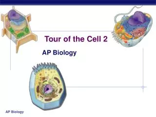
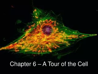
2. Eukaryotic cells contain a nucleus and organelles that are sub-cellular structures that perform specialized functions within the cell. Know the following from: Figure 6.9 (p. 100) – Overview of an animal cell. Figure 6.9 (p. 101) – Overview of a plant cell. a. Nucleus: contains majority of the genes in the eukaryotic cell present in the cells’ chromosomes. b. Cytoplasm: region between the nucleus and plasma membrane; organelles are suspended in this region c. Plasma Membrane: barrier that allows exchange of oxygen, nutrients, and waste into and out of the cell d. Cell Wall: maintains shape and structure of cell (found only in plant cells)
Animal Cell Figure 6.9
e. Ribosomes: carry out protein synthesis f. Endoplasmic Reticulum (ER): site of membrane synthesis and some metabolic processes. There are two types of ER: - Rough ER: covered with ribosomes (synthesis of proteins) - Smooth ER: surface lacks ribosomes g. Golgi Apparatus: modifies, stores, and secretes products of ER h. Lysosomes: bodies of enzymes that are used to digest macromolecules (found only in animal cells)and harmful compounds
i. Vacuoles: membrane-bounded sacs within cells - Food vacuoles: store and digest food - Contractile vacuoles: excrete excess water - Central vacuole: storage of excess organic and inorganic compounds; isolation j. Mitochondria: cellular respiration occurs and ATP is generated k. Chloroplasts: photosynthesis convert light energy to chemical energy (found only in plant cells)
Animal Cell Figure 6.9
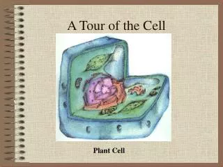
Plant Cell Figure 7.8
Further details and Figures: • The Nucleus (pp. 102 and 103) Note the double membrane and the chromatin (cellular DNA plus protein coat) in Fig. 6.10. • 2. Endomembrane System (pp 104 - 109). Functions to compartmentalize cellular activities. Includes the nuclear envelope, endoplasmic reticulum, Golgi apparatus, lysosomes, vacuoles, and the plasma membrane. In the film accompanying this section, note where a secretory protein is produced (endoplasmic reticulum, rough), how the protein moves to the Golgi apparatus (transported in a vesicle), that the protein is modified by the Golgi apparatus, and then transported to the plasma membrane where it is released to the outside of the cell. Note: secretory proteins are “secreted” to the outside of the cell so that they can perform functions outside the cell. Can you think of any?
3. Mitochondria and Chloroplasts Mitochondrion - site of respiration(production of energy from fats, sugars and other organic molecules resulting in carbon dioxide, water and waste
Chloroplasts - Site of photosynthesis (production of sugars and energy from carbon dioxide, water and light
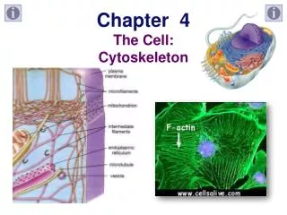
4. Cytoskeleton – Network of fibers that extend throughout the cytoplasm. Aids the cell in maintaining its shape. Especially true for animal cells that do not have cell walls, as plants cells do.Note Fig. 6.20 and Table 6.1. Remember the following two cytoskeleton components and their functions: Microtublues: subset of these responsible for the beating of cilia and flagella in eukarytoic cells. Note Figs. 6.23 and 6.24. Microfilaments: subset of these is responsible for the contraction of muscle cells. Note Fig. 6.27.