Download
1 / 37
370 likes | 400 Vues
Explore how chloroplasts convert solar energy and mitochondria harvest chemical energy. Learn about the evolution of eukaryotic cells from prokaryotes and the role of cytoskeletal structures. Understand the functions of cilia and flagella in cell movement.

E N D
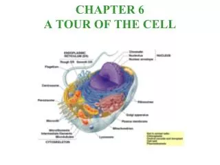
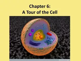
CHAPTER 4A Tour of the Cell Modules 4.15 – 4.21
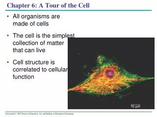
ENERGY-CONVERTING ORGANELLES 4.15 Chloroplasts convert solar energy to chemical energy • Chloroplasts are found in plants and some protists • Chloroplasts convert solar energy to chemical energy stored in sugars • The capturing of light and “electron energizing” occur in the grana and chemical reactions forming food storage molecules occur in the stroma. Chloroplast Stroma Inner and outer membranes Granum Intermembranespace Figure 4.15
4.16 Mitochondria harvest chemical energy from food • Mitochondria carry out cellular respiration • This process uses the chemical energy in food to make ATP for cellular work • ATP is the energy storage molecule. • Cellular respiration takes place in the presence of oxygen. • Carbon dioxide and water are given off by the cell during the oxidation of glucose. • ATP is produced for the cells energy needs
MITOCHONDRION Outermembrane Intermembranespace Innermembrane Cristae Matrix Figure 4.16
PROTISTS…the eukaryotes The eukaryotic cell probably originated as a community of prokaryotes—The Endosymbiotic Theory of Lynn Margulis (1970) • Eukaryotic cells evolved from prokaryotic cells more than 2 billion years ago • The nucleus and endomembrane system of eukaryotes probably evolved from infoldings of the plasma membrane of ancestral prokaryotes Plasma membrane Endoplasmic reticulum Nucleus Cytoplasm Nuclearenvelope Figure 16.18A Ancestral prokaryote Cell with nucleus and endomembrane system
Mitochondria and chloroplasts probably evolved from symbiotic prokaryotes that took up residence inside larger prokaryotic cells • Evidence comes from the fact that mitochondria and chloroplasts each have their own DNA that is similar to the DNA of bacteria. It also replicates independently of the nuclear DNA. Aerobic heterotrophicprokaryote Mitochondrion Mitochondrion Photosyntheticprokaryote Chloroplast Somecells Ancestral host cell Photosyntheticeukaryotic cell Figure 16.18B
THE CYTOSKELETON AND RELATED STRUCTURES 4.17 The cell’s internal skeleton helps organize its structure and activities • A network of protein fibers makes up the cytoskeleton Figure 4.17A
Tubulinsubunit Actin subunit Fibrous subunits 25 nm 7 nm 10 nm MICROFILAMENT INTERMEDIATEFILAMENT MICROTUBULE Figure 4.17B
Intermediate filaments reinforce the cell and anchor certain organelles • Microtubules • give the cell rigidity • provide anchors for organelles • act as tracks for organelle movement • Microfilaments of actin enable cells to change shape and move
A microfilament of actin is a globular structural protein that polymerizes in a helical fashion . These form the cytoskeleton - a three-dimensional network inside an eukaryoticcell. Actin filaments provide mechanical support for the cell, determine the cell shape, enable cell movements (through lamellipodia, filopodia, or pseudopodia); and participate in certain cell junctions, in cytoplasmic streaming and in contraction of the cell during cytokinesis..
The microfilaments are the thinnest component of the cytoskeleton, measuring only 5 nm in diameter. • Actin is one of the most abundant proteins in many eukaryotic cells, with concentrations of over 100 μM. It is also one of the most highly conserved proteins, differing by no more than 5% in species as diverse as algae and humans.
Intermediate filaments (IFs) are cytoskeletal structures formed by members of a family of related proteins. Intermediate filaments have a diameter between that of actin (microfilaments) and microtubules. Most types of intermediate filaments are located in the cytosol between the nuclear envelope and the cell surface membrane. Nuclear lamins are localized to the cell nucleus.
There are about 70 different genes coding for various intermediate filament proteins. However, different kinds of IFs share basic characteristics: they are all polymers that generally measure between 9-11 nm in diameter when fully assembled
Microtubules are protein structures found within cells, one of the components of the cytoskeleton. They have diameter of ~ 24 nm and length varying from several micrometers to possibly millimeters in axons of nerve cells. Microtubules serve as structural components within cells and are involved in many cellular processes including mitosis, cytokinesis, and vesicular transport. Microtubules are polymers of α- and β-tubulindimers
4.18 Cilia and flagella move when microtubules bend • Eukaryotic cilia and flagella are locomotor appendages that protrude from certain cells • A cilia or flagellum is composed of a core of microtubules wrapped in an extension of the plasma membrane • These structures are often associated with many mitochondria.
FLAGELLUM Electron micrograph of sections: Outer microtubule doublet Plasmamembrane Flagellum Centralmicrotubules Outer microtubule doublet Plasmamembrane Basal body Basal body(structurally identical to centriole) Figure 4.18A
Microtubule doublet • Clusters of microtubules drive the whipping action of these organelles Slidingforce Dynein arm Figure 4.18B
EUKARYOTIC CELL SURFACES AND JUNCTIONS 4.19 Cell surfaces protect, support, and join cells • Cells interact with their environments and each other via their surfaces • Plant cells are supported by rigid cell walls made largely of cellulose • They connect by plasmodesmata, channels that allow them to share water, food, and chemical messages
Walls of two adjacent plant cells Vacuole PLASMODESMATA Layers of one plant cell wall Cytoplasm Plasma membrane Figure 4.19A
Animal cells are embedded in an extracellular matrix of predominantly collagens. • It is a sticky layer of glycoproteins • It binds cells together in tissues like the mortar of a brick wall. • It can also provide a way of separating the tissues, and regulating intercellular communication • It can also have protective functions
Anchoring junctions link animal cells • Communicating junctions allow substances to flow from cell to cell • Tight junctions can bind cells together into leakproof sheets. For this reason they are more superficial. TIGHTJUNCTION ANCHORING JUNCTION COMMUNICATING JUNCTION Plasma membranes ofadjacent cells Extracellularmatrix Figure 4.19B
4.20 Eukaryotic organelles comprise four functional categories • Eukaryotic organelles fall into four functional groups Table 4.20
Cell types and Their Morphology • At all levels of organization, biological structures are shaped by natural selection to maximize their ability to perform their functions. • Many cells have a structure that suits their function in the body. • There are about 210 different cell types in the human body.
The rods and cones of the retina SEM or TEM?
Columnar Epithelium such as that found in the intestine. > Surface area
2-4 pages …. Due December 14th or 15th • Choose one organelle or cellular body and discuss a disease which results if that structure is not functioning properly. • OR • Choose a cell type and explain how its structure is suited for its function. • References needed