Download
1 / 61
610 likes | 852 Vues
Chapter 4: Tour of the Cell. BIO100 Fall 2007. THE MICROSCOPIC WORLD OF CELLS. Cells must be tiny for materials to move in and out of them and fast enough to meet the cell’s metabolic needs. Organisms are either. Single-celled, such as most bacteria and protists

E N D
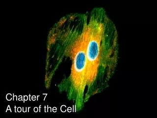
Chapter 4: Tour of the Cell BIO100 Fall 2007
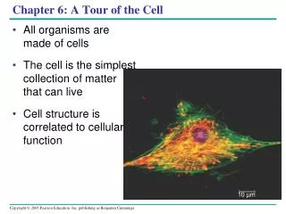
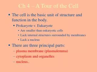
THE MICROSCOPIC WORLD OF CELLS • Cells must be tiny for materials to move in and out of them and fast enough to meet the cell’s metabolic needs.
Organisms are either • Single-celled, such as most bacteria and protists • Multicelled, such as plants, animals, and most fungi.
Microscopes as Windows to Cells • The light microscope is used by many scientists • Light passes through the specimen • Lenses enlarge, or magnify, the image. (a) Light micrograph (LM) of a white blood cell (stained purple) surrounded by red blood cells Figure 4.2A
How We Study Cells • Light Microscope: • First cells observed by Robert Hooke in 1665 using a light microscope. • Simple vs. Compound?
Magnification • An increase in the specimen’s apparent size • Resolving power • The ability of an optical instrument to show two objects as separate.
Cells were first discovered in 1665 by Robert Hooke • The accumulation of scientific evidence led to the cell theory, p. 57 • All living things are composed of cells • All cells form from previously existing cells • Cells are the smallest units capable of carrying out the processes of life: ex. respiration, digestion, reproduction, growth, ingestion, etc.
The electron microscope (EM) uses a beam of electrons • It has a higher resolving power than the light microscope.
Unaided eye • The electron microscope can magnify up to 100,000X Light microscope • Such power reveals the diverse parts within a cell. Electron microscope Figure 4.3
Metric Prefixes: Kilo=1000 so 10 Kcalories=10 000 calories= 10 C Hecto=100 Deka=10 Unit=1 ex. meter, liter, gram deci=0.1 centi=0.01 so 1 cm=10 mm milli=0.001
The scanning electron microscope (SEM) is used to study the detailed architecture of the surface of a cell. (b) Scanning electron micrograph (SEM) of a white blood cell Figure 4.2B
The transmission electron microscope (TEM) is useful for exploring the internal structure of a cell. (c) Transmission electron micrograph (TEM) of a white blood cell Figure 4.2C
The Two Major Categories of Cells • The countless cells on earth fall into two categories • Prokaryotic cells • Eukaryotic cells
Prokaryotic and eukaryotic cells differ in several respects. Prokaryotic cell Nucleoid region Eukaryotic cell Organelles Nucleus Figure 4.4
Are smaller than eukaryotic cells • Lack internal structures surrounded by membranes • Lack a nucleus. • Prokaryotic cells
Prokaryotic flagella Nucleoid region (DNA) Ribosomes Plasma membrane Cell wall Capsule Pili Figure 4.5
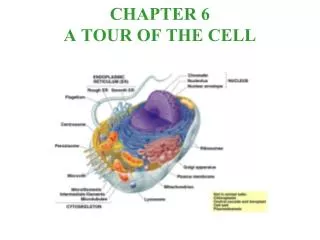
A Panoramic View of Eukaryotic Cells Centriole Ribosomes Not in most plant cells Lysosome Flagellum Cytoskeleton Plasma membrane Nucleus Mitochondrion Rough endoplasmic reticulum (ER) Smooth endoplasmic reticulum (ER) Golgi apparatus Figure 4.6A, p. 59
Not in animal cells Cytoskeleton • An idealized plant cell. Mitochondrion Central vacuole Nucleus Cell wall Rough endoplamsicreticulum (ER) Chloroplast Ribosomes Plasma membrane Smooth endoplasmic reticulum (ER) Plasmodesmata Golgi apparatus Figure 4.6B, p. 59
The nucleus is an organelle which contains long fibers made of DNA molecules and associated proteins. Each fiber, known as chromatin, becomes a chromosome Humans have 46 chromosomes in the nucleus of each and every cell Also within the nucleus is the nucleolus which is a ball-like mass of fibers and granules which produces the component parts of ribosomes.
Ribosomes move through the pores of the nucleus then are responsible for protein synthesis. Some are associated with “rough” ER others remain suspended in the cytosol.
MEMBRANE STRUCTURE AND FUNCTION • The plasma membrane separates the living cell from its nonliving surroundings • The entire region of cell between the nucleus and plasma membrane is the cytoplasm • Cytoplasm consists of organelles surrounded by a liquid known as cytosol.
A Fluid Mosaic of Lipids and Proteins • The membranes of cells are composed of • Lipids • Proteins.
Phospholipids form a two-layered membrane, the phospholipid bilayer. • The lipids belong to a special category called phospholipids Outside cell Hydrophilic head Hydrophobic tail Cytoplasm (inside cell) (a) Phospholipid bilayer of membrane Figure 4.7A
Most membranes have specific proteins embedded in the phospholipid bilayer. Hydrophilic region of protein Phospholipid bilayer Hydrophobic region of protein (b) Fluid mosaic model of membrane Figure 4.7B
Cytoplasm Fibers of extracellular matrix • Some functions of membrane proteins. c Enzymatic activity b Cell signaling a Attachment to cytoskeleton and extracellular matrix e Intercellular joining f Cell-cell recognition d Transport Cytoplasm Cytoskeleton Figure 4.8
Membrane phospholipids and proteins can drift about in the plane of the membrane • This behavior leads to the description of a membrane as a fluid mosaic • Molecules can move freely within the membrane • A diversity of proteins exists within the membrane.
Selective Permeability • Membranes of the cell are selectively permeable • They allow some substances to cross more easily than others • They block passage of some substances altogether.
The traffic of some substances can only occur through transport proteins • Glucose, for example, requires a transport protein to move it into the cell.
THE NUCLEUS AND RIBOSOMES:GENETIC CONTROL OF THE CELL • The nucleus is the manager of the cell • Genes found on the chromosomes within the nucleus store information necessary to produce proteins.
Structure and Function of the Nucleus • The nucleus is bordered by a double membrane called the nuclear envelope • It contains chromatin • It contains a nucleolus.
Ribosomes Chromatic Nucleolus Pore Nuclear envelope Figure 4.9
Ribosomes • Ribosomes build all the cell’s proteins.
How DNA Controls the Cell DNA 1 Synthesis of mRNA in the nucleus • DNA controls the cell by transferring its coded information into RNA mRNA Nucleus Cytoplasm mRNA 2 Movement of mRNA into cytoplasm via nuclear pore • The information in the RNA is used to make proteins. Ribosome 3 Synthesis of protein in the cytoplasm Protein Figure 4.10
THE ENDOMEMBRANE SYSTEM: MANUFACTURING AND DISTRIBUTING CELLULAR PRODUCTS • Many of the membranous organelles in the cell belong to the endomembrane system.
The Endoplasmic Reticulum Nuclear envelope • The endoplasmic reticulum (ER) Ribosomes • Produces an enormous variety of molecules • Is composed of smooth and rough ER. Rough ER Smooth ER Figure 4.11
Rough ER • Again, the “roughness” of the rough ER is due to ribosomes that stud the outside of the ER membrane.
The functions of the rough ER include • Producing proteins • Producing new membrane.
After the rough ER synthesizes a molecule it packages the molecule into transport vesicles 4 Transport vesicle buds off Secretory protein inside transport vesicle Ribosome 3 Protein 1 Rough ER 2 Polypeptide Figure 4.12
Smooth ER • The smooth ER lacks the surface ribosomes of ER and produces lipids, including steroids.
The Golgi Apparatus • Works in partnership with the ER • Refines, stores, and distributes the products of cells. Transport vesicle from ER “Receiving” side of Golgi apparatus Golgi apparatus New vesicle forming Transport vesicle from the Golgi “Shipping” side of Golgi apparatus Plasma membrane Figure 4.13
Lysosomes • A lysosome is a membrane-enclosed sac • It contains digestive enzymes • The enzymes break down macromolecules. So lysosomes are responsible for intracellular digestion. • If its membrane were to break its contents would digest the cell
They fuse with food vacuoles to digest the food. • Lysosomes have several types of digestive functions Lysosome Digestive enzymes Plasma membrane Digestion Food Food vacuole (a) Lysosome digesting food Figure 4.14a
They break down damaged organelles • They carry out the intracellular digestion. Lysosome Digestion Damaged organelle (b) Lysosome breaking down damaged organelle Figure 4.14b
Vacuoles • Two types are the contractile vacuoles of protists and the central vacuoles of plants. • Vacuoles are membranous sacs Central vacuole Contractile vacuoles (a) Contractile vacuoles in a protist (b) Central vacuole in a plant cell Figure 4.15
Rough ER Transport vesicle from ER Golgi apparatus • A review of the endomembrane system. Secretory vesicle from Golgi Vacuole Lysosome Secretory protein Plasma membrane Figure 4.16
CHLOROPLASTS AND MITOCHONDRIA: ENERGY CONVERSION • Cells require a constant energy supply to do all the work of life. • Nuclei, chloroplasts, and mitochondria are organelles having double membranes.
CHLOROPLASTS Inner and outer membranes of envelope • Chloroplasts are the sites of photosynthesis, the conversion of light energy to chemical energy. Granum Space between membranes Stroma (fluid in chloroplast) Figure 4.17
Mitochondria • Mitochondria are the sites of cellular respiration, which involves the production of ATP from food molecules. Outer membrane Inner membrane Cristae Matrix Space between membranes Figure 4.18
THE CYTOSKELETON:CELL SHAPE AND MOVEMENT • The cytoskeleton is an infrastructure of the cell consisting of a network of fibers.