Download
1 / 107
1.08k likes | 1.37k Vues
Chapter 6: A Tour of the Cell. Essential Knowledge. 2.a.2 – Organisms capture and store free energy for use in biological processes (6.2). 2.b.3 – Eukaryotic cells maintain internal membranes that partition the cell into specialized regions (6.2-6.5).

E N D
Essential Knowledge • 2.a.2 – Organisms capture and store free energy for use in biological processes (6.2). • 2.b.3 – Eukaryotic cells maintain internal membranes that partition the cell into specialized regions (6.2-6.5). • 4.a.2 – The structure and function of subcellular components, and their interactions, provide essential cellular processes (6.2-6.5). • 4.b.2 – Cooperative interactions within organisms promote efficiency in the use of energy and matter (6.4).
Light Microscope - LM • Uses visible light to illuminate the object • Relatively inexpensive type of microscope • Can examine live or dead objects • Light passes through specimen and then through various lenses • Lenses refract/bend light to magnify
Light Microscope Occular Lens Objective Lens Stage with specimen Light Source
Limitations - LM • Miss many cell structures that are beyond the magnification of the light microscope • Ex: lysosomes, centrioles • Need other ways to make the observations
Electron Microscopes • Use beams of electrons instead of light • Invented in 1939, but not used much until after WWII • Advantages: • Much higher magnifications • Magnifications of 50,000X or higher are possible. • Can get down to atomic level in some cases
Disadvantages of EM • Need a vacuum • Specimen must stop the electrons • High cost of equipment • Specimen preparation
Other Types of Microscopes • Transmission Electron Microscope - TEM • Sends electrons through thinly sliced and stained specimens • Gives high magnification of interior views. Many cells structures are now visible • Scanning Electron Microscopes – SEM • Excellent views of surfaces • Produces 3-D views • Live specimens possible
Limitations to EM • TEM: • Specimen dead; specimen prep is difficult • SEM: • Lower magnifications than the TEM; only see surface of specimen
TEM - interior SEM - surface
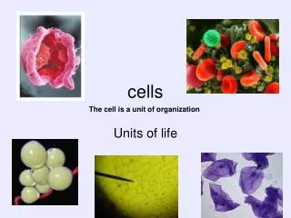
Cell Biology or Cytology • Cyto = cell - ology = study of • Should use observations from several types of microscopes to make a total picture of how a cell is put together • Directly related to biochemistry
Tools for Cytology • Cell Fractionation • Chromatography • Electrophoresis
Cell Fractionation • Disrupt cells • Separate parts (organelles and membrane) by centrifugation at different speeds • Separates by size and density of the various structures • Result - pure samples of cell structures for study
Chromatography • Technique for separating mixtures of chemicals • Separates chemicals by size or degree of attraction to the materials in the medium • Ex - paper, gas, column, thin-layer
Electrophoresis • Separates mixtures of chemicals by their movement in an electrical field • Used for proteins and DNA
Cell History • See alternate Ppt
History of Cells • Robert Hooke - Observed cells in cork • Coined the term "cells” in 1665 • Came from “jail cells” and/or monastery cells • Cells: • Al life is made of cells!!! • Cells are the simplest form of life
History of Cells • 1833 - Robert Brown, discovered the nucleus • 1838 - M.J. Schleiden, all plants are made of cells • 1839 - T. Schwann, all animals are made of cells. • 1840 - J.E. Purkinje, coined the term “protoplasm” • Late 1800s – Rudolf Virchow (“Omnis cellula e cellula” - All cells are from other cells)
Cell Theory: 3 Parts • All living matter is composed of one or more cells. • The cell is the structural and functional unit of life. • Cells come only from existing cells.
Two Types of Cells • 1) Prokaryotic- lack a nucleus and other membrane-bound structures. • 2) Eukaryotic- have a nucleus and other membrane-bound structures.
Eukaryotic Prokaryotic
Cell diversity • Most cells are between 5-50 micrometers • Mycoplasmas - bacteria that are .1 to 1.0 mm. (1/10 the size of regular bacteria) • # of cells: uni- and multicellular
Why Are Cells So Small? • Cell volume to surface area ratios favor small size • Nucleus to cytoplasm consideration (control) • Metabolic requirements
Surface area v. Volume • Vol and SA are proportionate (if one increases, the other increases) • Vol increases more than surface area (as cell grows) • Smaller objects have a greater ratio of sa to vol • Structure/Function: • Villi in intestinal cells – inc sa so cells can absorb more materials from food
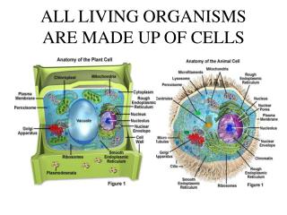
Basic Cell Organization • Membrane* • Nucleus • Cytoplasm* • Organelles • DNA/RNA* *EVERY cell has these 3 parts
Cell Membrane • Separates the cell from the environment • Boundary layer for regulating the movement of materials in/out of a cell • Often called plasma membrane • Bilayer of phospholipids • Allows oxygen, nutrients, wastes to pass through a series of processes: • Diffusion • Osmosis • Active transport
Cytoplasm • Cell substance between the cell membrane and the nucleus • The “fluid” part of a cell. • Neutral pH (serves as a natural buffer) • Exists in two forms: • gel - thick • sol - fluid
Organelles • Term means "small organ” • Formed body in a cell with a specialized function • Important in organizational structure of cells • More prominent/numerous in eukaryotic cells • Ex: Mitochondria, Endoplasmic reticulum, lysosomes
Nucleus • Most obvious organelle • Usually spherical, but can be lobed or irregular in shape • Contains genetic info • Found ONLY in euk cells • Function/s: • Control center for the cell • Contains the genetic instructions • Controls protein synthesis by making mRNA and rRNA (from DNA)
Structure of Nucleus • Nuclear membrane • Nuclear pores • Nucleolus • Chromatin
Nuclear Membrane • Otherwise known as Nuclear Envelope • Double membrane (lipid bilayer) separated by a 20-40 nm space • Inner membrane supported by a protein matrix (nuclear lamina) which gives the shape to the nucleus • Separates nuclear contents from cytoplasm • Dissolves during cell division
Nuclear Pores • Regular “holes” through both membranes • 100 nm in diameter • Protein complex gives shape • Lines every nuclear pore • Allows materials, such as macromolecules, in/out of nucleus
Nucleolus • Dark staining area in the nucleus • 0 - 4 per nucleus • Storage area for ribosomes • rRNA made here (from DNA) • No membrane encloses it??? (Research about nucleolus continues!!!)
Chromatin • Chrom: colored - tin: threads • DNA and protein in a “loose” format • Will form the chromosomes during Interphase of cell division (Chromosomes more condensed) • Each eukary cell has specific #
Ribosomes • Structure: 2 subunits made of protein and rRNA • No membrane • Function: protein synthesis • The more occurrences of protein synthesis, the more ribosomes • Ex: Pancreatic cells have over 1.2 million ribosomes
Ribosome structure • 2 Subunits: • 1) Large • 45 proteins, 3 rRNA molecules • 2) Small • 23 proteins, 1 rRNAmolecule • 2 Locations: • 1) Free in the cytoplasm- make proteins for use in cytosol • 2) Membrane bound- make proteins that are exported from the cell (Attached to rough ER)
Endomembrane System • Series of membranes connected by direct physical continuity or by transfer of membrane segments called vesicles • Includes: ER, Golgi, vesicles • Function: protein synthesis, transport of proteins, move lipids, detoxify proteins • Works closely with: nucleus, lysosomes, ribosomes, plasma membrane