Download
1 / 73
730 likes | 768 Vues
Explore the classification, etiology, presentation, diagnostic tests, treatment, follow-up, and complications of acute sinusitis. Learn about predisposing conditions, clinical presentations, pain patterns, and signs of dangerous complications.

E N D
Acute Sinusitis Diagnosis, Management, and Complications Jim Holliman, M.D., F.A.C.E.P. Professor of Military and Emergency Medicine Uniformed Services University of the Health Sciences Clinical Professor of Emergency Medicine George Washington University Bethesda, Maryland, U.S.A.
Acute Sinusitis Lecture Outline • Classification • Etiology • Presentation • Diagnostic tests • Treatment • Follow-up • Complications
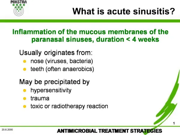
Sinusitis Classification • Definitions • Acute • Sx & signs of infectious process < 3 weeks duration • Subacute • Sx & signs 21 to 60 days • Chronic • > 60 days of sx & signs • Or, 4 episodes of acute sinusitis each > 10 days in a single year
General Contributors to Chronic Sinusitis • Resistant infectious organisms • Underlying systemic illness (esp. diabetes) • Immunodeficiency • Irreversible mucosal changes • Anatomic abnormality
Sinusitis Incidence • Reportedly > 31 million cases in U.S. • ? most common chronic illness • Is in 17 % of patients > age 65 • May occur in 0.5 to 1.0 % of all URI's
Sinusitis Pathogenesis • Basic cause is osteomeatal complex (the middle meatal region & the frontal, ethmoid, & maxillary sinus ostia there) inflammation & infection • Sinus ostia occluded • Colonizing bacteria replicate • Ciliary dysfunction • Mucosal edema • Lowered PO2 & pH
Development of the maxillary sinus (numbers are age in years)
Sinusitis Etiologic Organisms (& % incidence) • Aerobic bacteria • Strep. pneumoniae (30) • Alpha & beta hemolytic Strep (5) • Staph. aureus (5) • Branhamella catarrhalis (15 to 20) • Hemophilus influenzae (25 to 30) • Escherichia coli (5) • Anerobes (10 % acute, 66 % chronic) • Peptostreptococcus, Propionobacterium, Bacteroides, Fusobacterium • Fungi (2 to 5) • Viruses (5 to 10)
Acute Sinusitis Predisposing Conditions • Local • URI • Allergic rhinitis • Nasal septal defects • Barotrauma (diving) • Nasal foreign bodies • Nasal tubes • Dental infections • Overuse of topical decongestants • Nasal polyps or tumors • Aspiration of infected water • Smoking
Acute Sinusitis Predisposing Conditions (cont.) • Systemic • Diabetes • Immunocompromise (AIDS) • Malnutrition • Blood dyscrasias • Cystic fibrosis • Chemotherapy • Long term steroid Rx
Normal Functions of the Components of the Sinuses • Ostia • Drain secretions from sinuses • Allow pressure equalization • Diameter 2 to 5 mm (maxillary), 1 mm (ethmoid) • Cilia • Beat at frequency 1000 strokes/min. toward ostia • Push secretions out of sinus • Sinus secretions • 2 layered mucus • Contain IgA & IgG • Patency of ostiomeatal complex required for sinusitis resolution
Acute Sinusitis Usual Clinical Presentation • Symptoms progress over 2 to 3 days • Nasal congestion & discharge (usually thick & colored, not clear) • Localized pain +/- referred pain • Tenderness or pressure sensation over sinuses • Headache • Cough due to postnasal drip • Halitosis • Malaise
Usual Physical Findings With Acute Sinusitis • Erythematous edematous nasal mucosa • Purulent secretions in middle meatal area • May be absent if ostia completely blocked • Percussion tenderness • Over the involved sinuses • Over the maxillary molar +/- premolar teeth • Halitosis • +/- fever
Pain Patterns with Acute Sinusitis • Maxillary sinusitis • Unilateral pain over cheekbone • Maxillary toothache • Periorbital pain • Temporal headache • Pain worse if head upright • Pain better if head supine
Pain Patterns with Acute Sinusitis (cont.) • Ethmoid sinusitis • Medial canthal pain • Medial periorbital or temporal headache • Pain worsened by Valsalva or if supine • Sphenoiditis • Retroorbital, temporal, or vertical headache • Often deep seated headache with multiple foci • Pain worse supine or bending forward • Frontal • Frontal headache • Pain worse supine
Signs of Potentially Dangerous Complications of Acute Sinusitis • Periorbital, frontal, or cheek edema • Proptosis • Ophthalmoplegia • Ptosis • Diplopia • Meningeal signs • Neuro deficits of cranial nerves II to VI
Acute Sinusitis Use of Cultures • Routine culture of nasal secretions not useful • Poor correlation between non-directed nasal or nasopharyngeal culture isolates & sinus aspirate cultures • Sinus aspirate cultures useful only for protracted or nonresponsive sinusitis • Require endoscopy or needle puncture of sinus
Use of Paranasal Sinus Transillumination to Diagnose Sinusitis • First remove patient's dentures • Use darkened room • Shield light source from observer's eyes • Use Welch Allyn transilluminator or Mini-Mag Lite • Shine light over max. sinus & observe light transmission thru hard palate • Report results as opaque, dull, or normal for either side • Not useful for frontal sinuses since they often have developed asymmetrically
Sensitivity of Transillumination to Diagnose Sinusitis • Different studies have reached opposite conclusions on its usefulness ("Highly predictive" versus "criminal negligence") • Some studies have indicated it is useful if sinus is completely opaque (c/w Dx of sinusitis) or is completely normal (c/w absence of sinusitis), but has poor predictive value & correlation if transmission is "dull" • Can't be done in about 25 % of children due to poor cooperation
Acute Sinusitis Radiography • Plain films not as sensitive as CT • Radiographic signs of sinus pathology : • Air fluid levels • Partial or complete opacification • Bony wall displacement • 4 mm or more of mucosal wall thickening • Single Water's view has high concordance with 4 view sinus series (Caldwell, Water's, lateral, & submental vertex views)
Water’s view showing air-fluid level in right maxillary sinus and mucosal thickening in left maxillary sinus
Hypoplastic left frontal sinus and nosocomial right maxillary sinusitis
Limitations of Plain Film Radiography for Sinusitis • Poor visualization of ethmoid air cells • Difficulty distinguishing between infection, tumor, or polyp if sinus is completely opacified
Use of Ultrasound for Diagnosis of Sinusitis • Less sensitive than 4 view X-ray • Shown to not correlate well with sinus cultures • Accuracy is operator dependent • CT preferred for evaluation of complications
Another diagnostic modality for sinusitis is nasal endoscopy
Nasal endoscopic view showing uncinate process (U) displaced against middle turbinate (T) & closed off opening to frontal recess (arrow) from acute sinusitis
Nasal endoscopic view showing Aspergillus fungal mass arising from the sphenoid sinus
Use of Computed Tomography (CT) for Diagnosis of Sinusitis • Advantages of CT : • Visualizes ethmoid air cells • Evaluates cause of opacified sinus • Differentiates bony changes of chronic inflammation from osteomyelitis • Indicated only if complications suspected or if diagnosis uncertain (not needed initially for most cases)
CT scan showing fluid with pockets of air in frontal air cells from frontal sinusitis in a six year old male
Infectious and Granulomatous Diagnoses to Consider in the Differential Diagnosis of Sinusitis • Nasopharyngitis / adenoiditis • Dental abscess • Vestibulitis / furunculosis • Sarcoidosis • Tuberculosis • Rhinosporidiosis • Syphilis • Leprosy • Wegener's Granulomatosis • Midline (lethal) granuloma • Nasopharyngeal cancer
Lab Work for Diagnosis of Acute Sinusitis • Not helpful !
Goals of Medical Therapy for Acute Sinusitis • Control Infection • Facilitate sinus ostial patency and drainage • Provide relief of symptoms • Evaluate and treat any predisposing conditions to prevent recurrences
General Treatment for Acute Sinusitis • Oral antibiotic • Topical and systemic decongestants • Pain medications • Optional or secondary medications: • Guaifenesin (1200 mg po q 12h) • warm nasal saline irrigations qid • Antihistamine orally : only in the small % of patients with true allergic component
First - Line Antibiotic Therapy for Acute Sinusitis • Treatment duration should be 10 to 14 days (one recent study indicated 3 days may be OK) • Amoxicillin 500 mg po q 8 h • Augmentin 500 mg po q 8 h • Trimethoprim / Sulfamethoxazole DS one po bid • Azithromycin 500 mg po then 250 mg po q d x4 • Pediazole (Erythromycin - sulfisoxazole) QID may be best choice in kids