Download
1 / 54
650 likes | 1k Vues
Epidemiology of Osteoporosis. By : P.MOTTAGHI MD Associate Professor of Medicine – Rheumatology Department. Bone accretion occurs during adolescence, when there is a large increment in bone mass. Peak bone density is normally achieved after puberty and into the third decade of life.

E N D
Epidemiology of Osteoporosis By : P.MOTTAGHI MD Associate Professor of Medicine – Rheumatology Department
Bone accretion occurs during adolescence, when there is a large increment in bone mass. • Peak bone density is normally achieved after puberty and into the third decade of life. • However, by age 22, most individuals have achieved their peak bone mass. • At menopause, an acceleration of bone loss usually occurs over approximately 5 to 8 years, with an annual 2% to 3% loss of trabecular bone and a 1% to 2% loss of cortical bone. • Both men and women lose bone with age. Over a lifetime, women lose approximately 50% of trabecular and 30% of cortical bone; men generally lose two thirds of these amounts.
Genetics, gender, and racial differences • There are few data on ratio or gender differences in age related bone loss ,although limited evidence suggests that bone loss at the hip is greater in women than in men. • Lifestyle factors and level of economic development also contributes to the geographic variability in facture risk. • Peak bone mass determined largely by genetics . • Genetic factors account for about 70 to 80% of interdividual variation in peak bone mass in both gender. • Environmental factors , especially diet and mechanical loading ,also play a role in determining peak bone mass . • There are few data on racial or gender differences in age- related bone loss .
BONE REMODELING • Brief summery animation
Osteoporosis was previously thought to be a silent disease that was part of the normal aging process. • However, the advent of bone densitometry has made it possible to accurately and reproducibly identify patients at risk for osteoporosis so that prevention and treatment strategies can be instituted to reduce fractures.
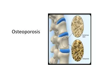
Osteoporosis, the most common metabolic bone disease, affects 200 million individuals worldwide. • Osteoporosis, or “porous bone,” is a “disease characterized by low bone mass and structural deterioration of bone tissue, leading to bone fragility and an increased susceptibility to fractures, especially of the hip, spine and wrist. • Initial studies of the epidemiology of osteoporosis was based on bone mineral content ,but no international agreement on the interpretation of the results made. Some groups rely on age- and sex-specific ‘normal’ ranges; others interpret their data in the light of the biomechanical concept of an absolute fracture threshold. • Therefore, the epidemiology of osteoporosis is still predominantly identical with the epidemiology of its major consequence, i.e. certain types of bone fractures claimed to be associated with osteoporosis
Epidemiology • 40% of women over 50 have osteopenia 7% of women over 50 have osteoporosis • Presence of osteoporosis carries 4-fold increase in fracture rate (over 50 years old) • Among those who live to 90 years old, 1/3 of women and 1/6 of men will have sustained osteoporotic fracture
After 50 years of age, there is an exponential rise in fractures, such that 40% of women and 13% of men develop one or more osteoporotic fractures in their lifetimes. • In the United States alone, there are more than 1.5 million osteoporotic fractures annually, including 250,000 hip, 250,000 wrist, and 500,000 vertebral fractures. • Hip fractures are associated with a 12% to 24% mortality rate in women and a 30% mortality rate in men within the first year of fracture, and 50% of patients are unable to ambulate independently and require long-term nursing home care. • These numbers will continue to grow exponentially as the elderly population of industrialized nations increases.
Clinical Crisis • 25 million women with osteoporosis or osteopenia in US in 2003 • At 50 years, 10% population • At 65 years, 20-25% population • At 75 year, 40% population • $13 billion for care of patients with osteoporotic spinal fracture in 1997 Vaccaro 2003
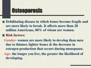
Risk factors for osteoporosis • Female gender • Age (post-menopausal or > 70 years) • Asian or Caucasian • Smoking, alcohol consumption • Thin body shape • Inactivity/immobility • Diet low calcium, high protein, caffeine, sodium • Some drugs glucocorticoids, chemotherapy, etc. • Some systemic diseases gastrectomy, chronic liver or renal disease, etc.
Risk Factors Female Gender 3X more likely to have hip or vertebral fracture than men 6X more likely to have forearm fracture Caucasian Race Higher than African-American, Asian race Smoking Low Body Weight (less than 58 kilos)
Risk Factors (cont’d) Sedentary Lifestyle Excessive Alcohol Intake Ample suggestion that moderate alcohol intake may be protective No clear threshold Nursing Home Residents 10X more likely to experience hip fracture than age-matched non-residents
Predisposing Medical Conditions Estrogen Deficiency Inflammatory Bowel Disease Type 2 Diabetes Mellitus Celiac disease Cystic fibrosis Hyperthyroidism Hyperparathyroidism Hypogonadism Liver Disease Corticosteroid use Heparin use Cyclosporine use Depo-Provera use
Risk Factors (cont’d) No clear increase in risk with carbonated beverages Although unclear risk association with excessive caffeine
Bone mineral density and fracture • The relationship between BMD and osteoporosis can be compared with that between blood pressure and stroke. • Although low BMD is not a prerequisite for osteoporotic fracture, the risk for fracture is elevated considerably in the presence of low bone mass. • Therefore, as with blood pressure, appropriate cut-off values can be defined to direct intervention toward ‘‘at-risk’’ individuals. • BMD taken at different sites can be used to predict the future risk for fracture at the same, or other, sites
Bone Mineral Density (BMD) and fracture rate Siris et al. Arch Intern Med. 2004; 164:1108-1112
BMD does not fully explain increased fracture risk in the elderly Age-related changes in bone quality are not fully captured by BMD Bone structure Material properties of bone Increased risk of falls
BMD Does Not Fully Explain the Effect of Age on Fracture Risk Fracture risk BMD 50 80 Age
Determinants of Bone quality (1) Bone Structure Macroarchitecture Microarchitecture Cross sectional diameter Hip axis length Cortical Porosity Trabecular Perforation
Morbidity • Overall, a 50-year-old white woman in the United States has a 13% chance of experiencing attributable functional decline after any fracture. • The degree of functional recovery after this injury is age dependent; in the United States, 14% of patients in the 50- to 55-year age group who sustained a hip fracture were discharged to nursing homes, compared with 55% of those aged older than 90 years. • Hip fracture also has a significant effect on mobility; 1 year after hip fracture, 40% were unable to walk independently, 60% required assistance with at least one essential activity of daily living (eg, dressing, bathing), and 80%were unable to perform at least one instrumental activity of daily living (eg, driving, shopping)
The impact of a single vertebral fracture may be low; however, multiple fractures cause progressive loss of height and kyphosis and severe back pain in the acute stages. The resultant loss of mobility can exacerbate underlying osteoporosis, which leads to the increased risk for further fractures. • Although good functional recovery after distal forearm fracture may be poor, reflecting complications (eg, reflex sympathetic dystrophy, neuropathies, posttraumatic arthritis), mortality after Colles’ fracture does not deviate from the expected rate.
Clinical manifestations • Fracture • Spine • Hip • wrist
Clinical manifestations Height loss Kyphosis
Symptoms • Early • Loss of bone mineral density • Late • Spine (“Dowager Hump”) & Fx • Hips Fx • Colles’ (wrist) Fx • Secondary Affects • Depression • Pain • Deformity • Dependency • Fear of falling • Premature death
Fracture Incidence Melton 1995
Hip Fracture • Most commonly treated fracture with respect to osteoporosis • Requires surgical intervention for future ambulation • Risk of morbidity 5-20% increase • The highest rates of hip fracture are seen in white populations that live in northern Europe, where the age-adjusted 1-year cumulative incidence in Norway in 1989 was 903/100,000 for women and 384/100,000 for men. • The rates are intermediate in Asians, in China, and in Kuwait, and are lowest in black populations. • In Western populations, among individuals who are older than 50 years of age, there is a female preponderance of hip fracture, with a female/male incidence ratio of approximately 2:1.
Risk Factors for Hip Fracture Largely Independent of BMD Previous fracture Family history Glucocorticoid therapy Smoking Alcohol intake Rheumatoid arthritis
Seasonal Variations in Vitamin D and Hip Fracture (Paso et al, 2005, JBMR 19:752)
vertebral Fractures • Thoracic • Height loss (often of several inches) with multiple sites, kyphosis, and secondary pain, discomfort related to altered biomechanics of the back, restricted respiratory disease • Lumbar • Abdominal symptoms including distention, early satiety, and constipation • 60% of vertebral fractures remain undiagnosed during first year of occurrence. • Only about one third of all vertebral deformities that are noted on radiographs come to medical attention, and less than 10% necessitate admission to the hospital
Risk Factors for Vertebral Fracture Age Gender Previous fracture Low BMD Premature menopause Smoking Use of a walking aid
Recent data from the Epidemiology of Osteoporosis Study yielded estimates of the prevalence of vertebral fractures to be 19% among women aged 75 to 79 years, 21.9% among women aged 80 to 84 years, and 41.4% among those aged 85 years and older. • Only one quarter of vertebral fractures result from falls. Most are precipitated by routine daily activities (eg, bending or lifting light objects),
Wrist fracture • In white women, the incidence increases linearly between the ages of 40 and 65 years, and then stabilizes. • There is no apparent increase in the incidence of wrist fracture with age in men.; in men, the incidence remains constant between 20 and 80 years. • United Kingdom showed that among women, the incidence of distal radius fracture increased from a premenopausal baseline of 10 per 10,000 population per year to a peak of 120 per 10,000 population per year over 85 years.
Other fractures • The incidence of proximal humeral, pelvic, and proximal tibial fractures also increase steeply with age, and are greater in women than in men.
Osteoporotic fracture: Influence of Age and Gender Hip Vertebrae 4000 Colles’ 3000 2000 1000 20 35–39 85 Men Women Incidence/100,000 person-years 35–39 85 Age group (years)