Download
1 / 47
480 likes | 568 Vues
Overview Of Osteoporosis. OBJECTIVES. Understanding the definition of osteoporosis Causes of osteoporosis Impact of osteoporosis Diagnosis of osteoporosis Treatment of osteoporosis. Bone has three major functions:.

E N D
OBJECTIVES • Understanding the definition of osteoporosis • Causes of osteoporosis • Impact of osteoporosis • Diagnosis of osteoporosis • Treatment of osteoporosis
Bone has three major functions: • Provide rigid support to extrimities and body cavities containing vital organs. • Provide efficient levers and sites of attachment of muscles which are all crucial to locomotion. • Provide a large reservoir of ions such as calcium, phosphorus, magnesium and sodium which are critical for life and can be mobilized when the external environment fails to provide them
I. Osteoblasts: The bone forming cells which are actively involved in the synthesis of the matrix component of bone (primarily collagen) and probably facilitate the movement of minerals ions between extracellular fluids and bone surfaces. II. Osteocytes: The are believed to act as a cellular syncytium that permits translocation of mineral in and out of regions of bone removed from surfaces. III. Osteoclasts: The bone resorption cells.
OSTEOPOROSIS THE DISEASE OF THE 21ST CENTURY Lancet ,MAY,2002 By Stephanie Clark
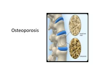
Definition of osteoporosis Skeletal disorder chracterized by compromized bone strength predisposing a person to an increased risk of fracture. Bone strength reflects the integration of bone density and bone quality
Definition Decrease in bone mass and strength associated with an increased tendency to fractures
Type I Osteoporosis(Post Menopausal) Fractures of bones composed mainly of Trabecular bone. e.g., Distal Radius - Colle’s fracture Vertebra - Crush & Wedge fractures Usually affects woman within 15 years of menopause.
Type II Osteoporosis(Senile) Fractures of bones composed of both cortical & Trabecular bone. e.g., Hip - Femure neck fracture Usually affects individual over age of 70 years.
Risk Factors: non-modifiable • Age (increasing) • Low BMI (small, low weight;< 58 kg) • Ethnicity: Caucasian > Asian/Latino > African American • Family History of Fracture
Risk Factors: Modifiable • Sex Hormones (low estrogen/testosterone) • Low calcium and vitamin D • Inactive lifestyle • Excessive alcohol • Cigarette smoking • Rheumatoid arthritis • Hyperparathyroidism (primary or secondary) • Hyperthyroidism • GI conditions which impair adequate nutrition • Steroids or Cushing’s • Proton pump inhibitors
Secondary Factors causing Bone Loss Factors Associated with Decreased Bone desity
Clinical presentation of osteoporosis • Generally patients are asymptomatic even with very low bone densities • Hip Fractures • Acute or chronic back pain secondary to vertebral fractures • Atraumatic or low impact fractures
Laboratory & Radiological Findings Bone profile ,ALP and PTH are within normal in patients with osteoporosis due to sex hormones deficiency and aging. X-rays of skeleton do not show a decrease in osseous density until at least 30% of bone mass has been lost.
WHO 1994 Definition based on BMD : Normal : greater than or equal to -1 SD Osteopenia: BMD which lies between - 1 and -2.5 SD Osteoporosis : less than or equal to – 2.5 SD Severe osteoporosis : osteoporosis with 1 or more fragility fractures
Younger individuals • USE Z SCORE (comparison to age-matched norms) • If ≤ 2 ( below expected range for age)
Hip fractures are bad • 20% patients with hip fracture die within the year • 25-30% need placement in skilled nursing facility
Epidemiology of fracturesVertebral fractures Affected Vertebral fractures : rarely reported by physicians 10 % of vertebral fractures result in hospitalizations Prevalence increases with age Male to female ratio 1: 1 Mid thoracolumbar region are most commonly affected. Cause lower energy,poor slep,pain,immobility and social isolation especially in men. Back deoformities :loss of height and kyphosis.
Hip fractures Cause serious disability and excess mortality Highest incidence in Scandinavian and N American countries. Women who have sustained fracture have a 10-20 % higher mortality than would be expected for their age. Above 50 years of age , female to male ratio is 2: 1. Mortality is higher in men , greater with co existent diseases 1-year mortality : 31 % in men and 17% in women Risk of death is greatest immediately post fracture
Economic Impact Huge Osteoporotic fractures cost the US 17.9 billion per annum UK : 1.7 billion Cost is largely attributed to hip fractures
Estimated number of hip fractures 1990-2025-2050Cooper et al,2008
WHEN TO SCREEN WITH DXA SCAN • VERY CONTROVERSIAL • IN US AND CANADA : WOMEN≥ 65 YEARS • MEN≥ 70 YEARS • SCREEN IN INDIVIDUALS WITH RISK FACTORS EG. STEROIDS • EUROPE : CASE FINDING IE IN PEOPLE WITH RISK FACTORS
Osteomalacia Failure of organic matrix (osteoid) of bone to mineralize normally. Commonest cause is vitamin D deficiency.
Exclude secondary causes especially in younger individuals and men
Prevention • Adequate nutrition, particularly calcium and vitamin D • Calcium: 1000 – 1200 mg daily (diet plus supplementation) • Vitamin D: goal level above 50-75 nmol/l • Weight bearing exercise • Discourage smoking • Reduction of risks for falling: consider OT evaluation for home hazards, minimize sedating medications. • Hip protectors: can be useful if worn properly but often have low compliance
Treatment The Adolescent Group(Peak bone mass attainment) “Senile Osteoporosis is a peadiatric disease”. • A calcium intake of 1200 mg/day is recommended. • Adequate sun exposure or vit D supplementation to ensure adequate level. • A reasonable exercise program is recommended. • Genetic influence on peak bone mass attainment.
MANAGEMENT • Nonpharmacologic • Modification of life style measures • Exercise • Prevention of falls • Adequate calcium and vitamin D intake • Stopping smoking • More sun exposure
Calcium and Vitamin D • At least 1000 mg /day for men ≤ 65 or younger • 1500 mg /day for older men. Ca citrate vs. Ca carbonate. • Vitamin D : check 25 (OH) vit. D level . If very low you need to “replete” the stores first . Maintenance dose is 800 IU for men younger than 50 and 800-1000 IU for men older than 50 • 1000 IU or more for all patients with osteoporosis or reduced bone mass regardless of their age.
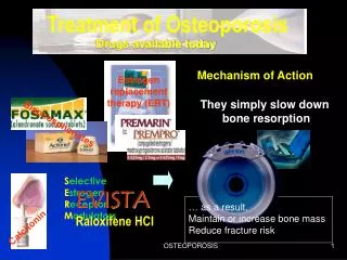
Treatment Options • . Bisphosphonates • 2. Denosumab • 3. Teriperatide • 4. SERMs (Selective estrogen receptor modulators) – • 5. Hormone replacement therapy • 6. Calcitonin : no longer used
summary • . Screening • All women > 65 years • Men > 70 • Women 50-64 with risk factors • Patients on steroids or anti-estrogen/anti-testosterone treatment • 2. Prevention with adequate calcium/vitamin D, weight bearing exercise should be advised for all. • 3. DXA scan is the primary screening tool • 4. Aggressive therapy should be offered to patients with atraumatic/low-impact fractures and those with osteoporosis, osteopenia with mulitple risk factors, patients on steroids, anti-estrogen, and anti-testosterone therapy with abnormal bone densities (T score <-1).