Blood Coagulation
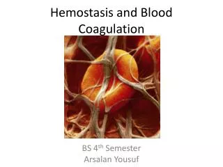
Blood Coagulation. Haemostasis part2. ROLE OF ENDOTHILIUM . Hemostasis:. BV Injury. Tissue Factor. Neural. Coagulation Activation. Blood Vessel Constriction. Platelet Activation. Primary hemostatic plug. Reduced Blood flow. Plt-Fusion. Thromibn, Fibrin. Stable Hemostatic Plug.

Blood Coagulation
E N D
Presentation Transcript
Hemostasis: BV Injury Tissue Factor Neural Coagulation Activation Blood Vessel Constriction Platelet Activation Primary hemostatic plug Reduced Blood flow Plt-Fusion Thromibn, Fibrin Stable Hemostatic Plug
Coagulation: Intrinsic 12,11,9,8 (aPTT-) Extrinsic-7 (PT) Common Path (TT) FX FXa Prothrombin Thrombin Fibrinogen Fibrin
Clot formation & retraction Fibrinogen Thrombin F-XIIIa Fibrin Mononer Fibrin Polymer Cross Linked Fibrin
Profibrinolytic Plasminogen tPA Fibrin Fragment D-dimer Procoagulant Platelets Factors Fibrinogen von Willebrand Factor FORMATION RESOLUTION clot Antifibrinolytic PAI-1 Alpha-2 Antiplasmin Anticoagulant Antithrombin III Protein C Protein S
Coagulation Made Easy- The Mixing Study • Useful to differentiate etiologies of prolonged clotting in a coagulation assay. • Patient’s plasma is mixed 50:50 with normal plasma. Coagulation assay is repeated. • If “substantial” correction is noted after mix, suspect clotting factor deficiency. • If no or minimal correction seen, suspect inhibitor.
Disorders of the haemostatic mechanism are devided into three main groups: • Disorders of the vessels • Disorders of the platelets • Disorders of the coagulation mechanism („coagulopathies”) „purpuric diseases”
The investigation of a patient with a suspected disorder of haemostasis • case history (personal details, family history) • inspection (type of bleeding) • physical examination • other known diseases • drugs and medications • laboratory tests
Certain signs and symptoms are virtually diagnostic of disordered haemostasis. The main symptom of all diseases is the bleeding: ● in the „purpuric disorders” cutaneous and mucosal bleeding usually is prominent ● in different types of „coagulopathies” hemarthroses, haematomas are the characteristic bleeding manifestations. The onset of bleeding following trauma frequently is delayed (recur in a matter of hours) (the temporary hemostatic adequacy of the platelet plug may explain this phenomenon).
Petechiae, purpuras: small capillary haemorrhages ranging from the size of a pinhead to much larger
Haematomas: may be spontaneous (in a serious hemorrhagic disease) or may occur after trauma (in a mild hemorrhagic disease).
Intramuscular injection may be very dangerous to the patient with a bleeding disorder Venipuncture (if skilfully performed) is without danger becouse the elasticity of the venous walls.
Aquired:generally several coagulation abnormalities are present. Clinical picture is complicated by signs and symptoms of the underlying disease. Deficiencies of the vitamin K dependent coagulation factors (FII, VII, IX, X) Hepatic disorders Accelerated destruction of blood coagulation (DIC) Inhibitors of coagulation Others (massive transfusion, extracorporal circulation) Hereditary:deficiency or abnormality of a single coagulation factor. Hemofilia A (FVIII) Hemofilia B (FIX) Von Willebrand’s disease Rare coagulopathies (FI. II. V. VII. X. XI. XIII) Coagulopathies
Hereditary Factor DeficienciesHemophilia • x-linked recessive conditions (males only) • type A : F VIII:C deficiency (Classical Hemophilia) B : F IX deficiency (Christmas disease) C : F XI deficiency • unexplained bruising or bleeding in young males, usually ~ 1 yr of age • joint & muscle bleeding → arthropathy
Haemophilia A bleeding disorder in which clotting factor VIII (eight) /Haemophilia A/ or IX (nine) /Haemophilia B/ in a person's blood plasma is missing or is at a low level. Prevalence: haemophilia A: 105/million men haemophilia B: 28/million men
The hemophilia gene is carried on the X chromosome in males who lack a normal allele, the defect is manifested by clinical haemophilia. Women may be carriers.
Haemophilia is a lifelong disease • A person born with haemophilia will have it for life. • The level of factor VIII or IX in his blood usually stays the same throughout his life.
Hereditary Factor DeficienciesHemophilia ■ screening: prolonged PTT ■ hemophilia A mild moderate severe : life-threatening (CNS bleed) • treatment factor replacement rFVIIa
Hemophilia B(Christmas disease, Factor IX def.) • less common than hemophilia A • similar clinical Sx and inheritance pattern as hemophilia A (sex-linked) XII XI IX VIII VII X V II I XIII Stable clot
von Willebrand’s disease • easy bruisability (no bleeding into joints) • unable to release VIII-vWF • intact VIII-vWF synthesis • VIII-C level is also decreased (unknown reason) • autosomal dominant • 1 in 30,000 population • usually diagnosed in childhood or young adults • increased bleeding time • normal plt, PT • normal or increased aPTT
Hereditary Platelet Disordervon Willebrand Disease (vWD) • most common congenital bleeding disorder • quantitative or qualitative abn. of vWF • Type 1: most common form • partial quantitative deficiency of vWF • autosomal dominant • mucocutaneous bleeding • hematology consult prior to surgery • prolonged bleeding time, normal platelet
Hereditary Platelet Disordersvon Willebrand Disease (vWD) Type 2: qualitative alterations in the vWF structure & function Type 3: least common and most severe • Complete absence of vWF in plasma or storage organelle • Autosomal recessive • acquired vWD • Lymphoproliferative disease ▪ cardiac/valvular disease • Tumors ▪ medications (valproic acid) • Autoimmune disease ▪ hypothyroidism
Hereditary Platelet Disordersvon Willebrand Disease • Treatment: Desmopressin (DDAVP) • synthetic analog of vasopressin • ↑ both F VIII and vWF 3 - 5x in 30 mins • preop prophylactic dose: 0.3 mcg/kg IV in 50 -100 ml NS infused 30-60 mins q 12-24 hrs PRN • duration 8-10 hrs • intranasal dose: 300 mcg – for home treatment, not for preop prophylaxis
Hereditary Bleeding Disordersvon Willebrand Disease • DDAVP • vasomotor effect: flushing, ↑HR, headache • SE: hyponatremia, seizures • not for children < 3 yrs old • unresponsive to DDAVP (15%) • cryoprecipitate • FFP • factor VIII / vWF concentrate
Vitamin K Deficiencyvitamin K dependent factors : II, VII, IX, X • acquired disorder • may occur in malnutrition, malabsorption, biliary obstruction, drug • increased PT • normal bleeding time, plt • normal or increased aPTT XII XI IX VIII VII X V PT II I XIII Stable clot
Severe Liver Diseasefactors synthesized in liver : II, V, VII, IX, X, fibrinogen • increased PT, aPTT • normal bleeding time, plt XII XI IX VIII VII X V PT aPTT II I XIII Stable clot
Platelet Dysfunction, Factor Deficiencies & Presence of Inhibitors • Liver disease • synthesis of coagulation factors (except VIII), anticoagulants, ATIII, protein C & S, plasminogen • clearance of activated clotting factors, tPA, FDPs • DIC
Blood Component TherapyTransfusion of FFP 1) replacement of isolated factor deficiency 2) reversal of coumadin 3) antitrombin III deficiency 4) treatment of immunodeficiencies 5) treatment of TTP 6) massive blood transfusion
Blood Component TherapyCryoprecipitate • contains significant levels of factor VIIIC, factor VIII: vwF, XIII, fibrinogen • indications: 1) hemophilia 2) von Willebrand disease 3) fibrinogen deficiencies 4) uremic platelet dysfunction
Disseminated IntravascularCoagulation (DIC) - an acute, subacute, or chronic thrombohemorrhagic disorder occurring as a secondary complication in a variety of diseases - activation of clotting system resulting in wide spread formation of microthrombi throughout the microcirculation - as a consequence, causing consumption of platelets, fibrin and coagulation factors, and activation of thrombolytic mechanism Two major triggering mechanisms 1. release of tissue factor or thromboplastic substance 2. widespread endothelial injury
DIC Triggering Mechanisms 1. release of tissue factor or thromboplastic substance - placental tissue - granules from leukemic cells - bacterial endotoxin - mucus from adeno CA 2. widespread endothelial injury - Ag-Ab immune complex deposit - extreme temperature - microorganisms