Electrophoresis / SDS-PAGE
1.04k likes | 4.78k Vues
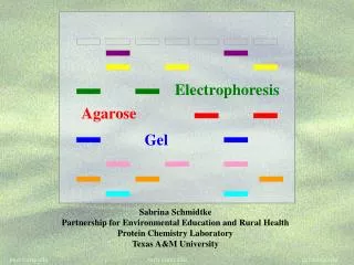
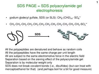
Electrophoresis / SDS-PAGE. Purpose. Use gel electrophoresis to compare protein profiles of fish. What is SDS-PAGE?. SDS (Sodium dodecyl sulphate) is a detergent used to denature proteins and give them a negative charge PAGE: Polyacrylamide Gel Electrophoresis

Electrophoresis / SDS-PAGE
E N D
Presentation Transcript
Purpose • Use gel electrophoresis to compare protein profiles of fish
What is SDS-PAGE? • SDS (Sodium dodecyl sulphate) is a detergent used to denature proteins and give them a negative charge • PAGE: Polyacrylamide Gel Electrophoresis • It is a technique to separate proteins by their molecular weight
Electrophoresis • The migration of proteins is affected by multiple factors involving their structural organization. • Amino acids can carry either a net positive, net negative, or neutral charge depending on the combination of amino acids they contain. • To make protein migration rates a function of molecular weight, it is necessary to impose a uniform shape and charge on all of the proteins in the mixture.
What is so special about SDS? • SDS is a negatively charged detergent. • Disrupts secondary and tertiary protein structures by breaking hydrogen bonds and unfolding protein. • ‘Masks’ charge on protein so that all proteins act the same as regards charge. • Prevents protein aggregation. • Prevents protein shape from influencing gel run.
- largest large small smallest + How does SDS-PAGE work? • Proteins (negatively charged due to SDS) move to positive electrode • Proteins separate by size • Smaller proteins move faster
s-s SDS, heat proteins with SDS - + Why heat the samples? • Heating the samples helps denature proteins and protein complexes, allowing the separation of individual proteins by size
Why Use Polyacrylamide Gels to Separate Proteins? • Smaller pore size than agarose • Proteins much smaller than DNA • average protein = 30-50 kD • “average” DNA = >2000 kD
Procedure: Day 1 • Label one flip-top and one screwcapmicrotube with the letter of each fish sample being prepared for electrophoresis. • Add 250µl of Laemmli Sample buffer to each labeled flip-top tube. • Transfer each fish sample to the appropriately labeled microtubeand close the lid. • Gently flick the microtubes 15 times with finger to agitate the tissue in the sample buffer. • Incubate samples for 5 minutes at room temperature. • Carefully pour the sample buffer containing the extracted proteins, but not the solid fish piece, into the correctly labeled screwcapmicrotube. • Heat the fish samples and the actin and myosin standard (AM) for 5 minutes at 95oC to denature the proteins. • Store samples in freezer until next class.
Procedure: Day 2 • Place a yellow sample loading guide on the top of the electrode assembly. • Load the samples into the wells following this guide:
Load 10 µl of each protein sample into the designated well. • After loading all samples, remove the sample loading guide, place the lid on the tank. • Run gel for 30 minutes at a constant voltage of 200V.
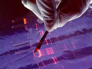
When the gels are finished running, discard the buffer from the inner chamber, release the cams, and remove the gel cassettes from the assembly. • Lay each gel cassette flat on the bench with the short plate facing up. Carefully pry apart the gel plates using your fingertips or a weighing spatula. The gel will adhere to one of the plates. • Transfer the plate with the gel to a staining tray containing Bio-Safe Coomassie stain. • Stain the gels for 1 hour. Gentle agitation throughout staining time gives best results. • After staining for 1 hour, pour off stain and return to bottle. • Destain the gels in a large volume of water, changing it several times if possible. Allow gels to destain overnight for best results.