Download
1 / 14
190 likes | 1.01k Vues
Agarose gel electrophoresis. BCH 333 [practical]. Agarose gel electrophoresis: is a method of gel electrophoresis used in biochemistry and molecular biology to separate and analyze DNA or RNA molecules by size. -It is used to determine the size of DNA, checking the purity of isolated DNA.

E N D
Agarose gel electrophoresis BCH 333 [practical]
Agarose gel electrophoresis: is a method of gel electrophoresis used in biochemistry and molecular biology to separate and analyze DNA or RNA molecules by size. -It is used to determine the size of DNA, checking the purity of isolated DNA. Principle: -Biomolecules [DNA or RNA] are separated by applying an electric field to move the negatively charged molecules [-] through an agarose matrix towards the anode [+] , and the biomolecules are separated by size in the agarose gel matrix. -The largest molecules will have the most difficulty passing through the gel pores, whereas the smallest molecules will move faster.
-After the samples of DNA are applied to the wells of agarose gel along with the tracking dye. -The power is turned on, after filling the buffer in the tank to allows current to pass. -The tracking dye and the DNA samples are negatively charged [-] and migrate towards the positive electrode [+]. -The DNA backbones contain negatively charged phosphate groups, which are attracted to the positive electrode. -Large DNA fragments retard earlier, while the smallest fragments move the fastest.
DNA: note the phosphate groups in the backbone are negatively charged.
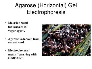
What is Agarose: -is a linear polymer of D-galactose and 3,6-anhydro-1-galactose and forms a gel that is held together by hydrogen bonds. - Agarose is present in powder forms that are dissolved in buffer close to boiling temperatures , then cooled down to around 40 degrees it sets and forms a gel . [polymerized] -The concentration of the material in the gel determines the size of the pores. [high concentration of the gel small pore size] . Agarose gels have larger pore sizes compared to Dextran and polyacrylamide gels. -This makes it useful for the analysis or separation of large globular proteins or long, linear molecules such as DNA. Prepare it ? Prepare agarose gel with 0.8%?
Buffer used: • -helps is deliver the electric current through the gel. • Buffer used is either TBE or TAE. • - TBE buffer: is made with Tris/Boric acid/ EDTA. • - TAE buffer: is made with Tris/Acetic acid/ EDTA. • Tracking Dye: • A dye such as bromophenolblue [or orange dye] is also included in the sample; • it makes it easier to see the sample that is being loaded and also acts as a marker of the electrophoresis front.
Gel staining: -The DNA in the gel needs to be stained and visualized, The reagent most widely used is the fluorescent dye ethidium bromide "EtBr“, that emits orange light after binding to DNA. Note: That the gel will be viewed under ultraviolet light. [Ethidium bromide is a cyclic planar molecule that binds between the stacked base-pairs of DNA.].
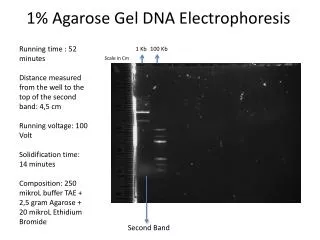
Determine the size of the DNA fragment: -Since agarose gels separate DNA according to size, the size of a DNA fragment may be determined from its electrophoretic mobility by running a number of standard DNA markers of known sizes on the same gel. Figure: The 100 bp DNA Ladder is suitable for sizing double-stranded DNA fragments from 100-2000 bp.
Note that: -The higher the voltage the more quickly the gel runs. [ Voltage rate of migration]. -Gel concentrations must be chosen to suit the size range of the molecules to be separated.
SDS-PAGE Vertical _ pace devices Running buffer Staining buffer [lamp] De-staining buffer Acrylamide gel [stacking, separation gel] Agarose gel electrophoresis Horizontal _ agarose_ devices Running buffer Stained and view using UV light Agarose gel