Download
1 / 11
130 likes | 750 Vues
Agarose gel electrophoresis. Agarose gel electrophoresis is an easy way to separate DNA fragments by their sizes and visualize them. It is a common diagnostic procedure used in molecular biological labs.

E N D
Agarose gel electrophoresis is an easy way to separate DNA fragments by their sizes and visualize them. It is a common diagnostic procedure used in molecular biological labs.
The technique of electrophoresis is based on the fact that DNA is negatively charged at neutral pH due to its phosphate backbone. For this reason, when an electrical potential is placed on the DNA it will move toward the positive pole:
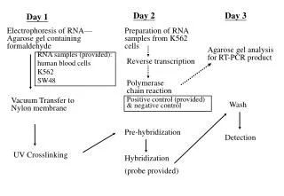
The equipment and supplies necessary for conducting agarose gel electrophoresis are relatively simple and include: • An electrophoresis chamber and power supply • Gel casting trays, which are available in a variety of sizes and composed of UV-transparent plastic. The open ends of the trays are closed with tape or with tank wall. • Sample combs, around which molten agarose is poured to form sample wells in the gel. • Electrophoresis buffer, usually Tris-acetate-EDTA (TAE) or Tris-borate-EDTA (TBE).
Loading buffer, which contains something dense (e.g. glycerol) to allow the sample to "fall" into the sample wells, and one or two tracking dyes, which migrate in the gel and allow visual monitoring or how far the electrophoresis has proceeded. • Ethidium bromide, a fluorescent dye used for staining nucleic acids. NOTE: Ethidium bromide is a known mutagen and should be handled as a hazardous chemical - wear gloves while handling. • Transilluminator (an ultraviolet lightbox), which is used to visualize ethidium bromide-stained DNA in gels. NOTE: always wear protective eyewear when observing DNA on a transilluminator to prevent damage to the eyes from UV light.
Preparation 1- Make a 1% agarose solution in 1x TBE. If you analyze small DNA strands, go up to 2%.
2- Boil solution, preferably in a microwave oven. 3- Let the solution cool down to about 50 °C at room temperature. Stir the solution while cooling. 4- Add 0.2ul ethidium bromide per 10 ml gel solution. Wear gloves from here on, ethidium bromide is a potent mutagen(nitrile gloves recommended) ! Some researchers prefer not to add ethidium bromide to the gel itself, instead soaking the gel in an ethidium bromide solution after running.
5- Stir the solution to disperse the ethidium bromide, then fill it into the gel rack. 6- Insert the comb at one side of the gel, about 5-10 mm from the border of the gel. 7- When the gel has cooled down and become solid, remove the comb. The holes that remain in the gel are the slots. 8- Put the gel, together with the rack, into a chamber with 0.5x TBE. Make sure the gel is completely covered with TBE, and that the slots are at the electrode that will have the negative current.
Factors affecting migration • The length of the DNA molecule • Increasing the agarose concentration of a gel reduces the migration speed and enables separation of smaller DNA molecules • The presence of ethidium bromide (EtBr) in the gel causes DNA to run slower • The higher the voltage, the faster DNA migrates (above about 5 to 8 V/cm).