Download
1 / 32
660 likes | 1.59k Vues
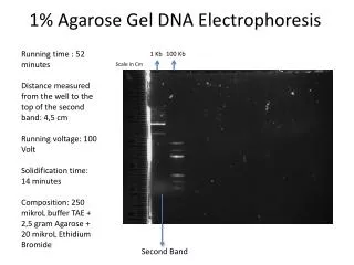
DNA Agarose Gel Electrophoresis. ( demonstration ). Electrophoresis. Principle is to separate DNA or proteins on the basis of their charge and their ability to migrate within a gel (jello-like) matrix . (molecular weight and structure)

E N D
DNA Agarose Gel Electrophoresis ( demonstration )
Electrophoresis Principle is to separate DNA or proteins on the basis of their charge and their ability to migrate within a gel (jello-like) matrix. (molecular weight and structure) A strong electric field is applied to DNA or the protein mixture for an extended period of time (hours) until the DNA move apart or migrate.
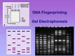
DNA small large - + Power • DNA is negatively charged. • When placed in an electrical field, DNA will migrate toward the positive pole (anode). How fast will the DNA migrate?
How fast will the DNA migrate? 1. strength of the electrical field 2. buffer (PH …..) 3. density of agarose gel… 4. Size of the DNA ! • gel electrophoresis separates DNA according to size • Small DNA move faster than large DNA • Within an agarose gel, linear DNA migrate inversely proportional to the log10 of their molecular weight.
Size of the DNA 1 )
The structure of plasmid DNA 2 ) cccDNA (covalently closed circular DNA) Oc DNA(open circular DNA) L DNA (linear DNA) Ccc DNA<L DNA <oc DNA
Step1: making the gel Buffer An agarose gel is prepared by combining agarose powder and a buffer solution. Flask for boiling Agarose
Electrophrosis buffer solution • 1) Tris-boratic acid(TBE) • 2) Tris-acetic acid(TAE) • 3) Tris-phosphate(TPE) 5×TBE: • Tris 108g • EDTA 9.3g • Boratic acid 55g • H2O to 1000ml pH : 8.0~8.2 Add H2O to 1×TBE if use
Agarose Buffer Solution Combine the agarose powder and buffer solution. Use a flask that is several times larger than the volume of buffer.
Melting the Agarose Agarose is insoluble at room temperature (left). The agarose solution is boiled until clear (right). Gently swirl the solution periodically when heating to allow all the grains of agarose to dissolve. ***Be careful when boiling - the agarose solution may become superheated and may boil violently if it has been heated too long in a microwave oven.
Electrophoresis Equipment Power supply Gel tank Cover Electrical leads Casting tray Gel combs
Gel casting tray & combs Preparing the Casting Tray Seal the edges of the casting tray and put in the combs. Place the casting tray on a level surface.
Pouring the gel Allow the agarose solution to cool slightly (~60ºC) and then carefully pour the melted agarose solution into the casting tray. Avoid air bubbles.
Each of the gel combs should be submerged in the melted agarose solution.
wells When cooled, the agarose polymerizes, forming a flexible gel. It should appear lighter in color when completely cooled (30-45 minutes). Carefully remove the combs and tape.
Step 2: Setting up the Apparatus DNA buffer wells Anode (positive) Cathode (negative) The agarose gel is placed in an electrophoresis apparatus. Add enough electrophoresis buffer to cover the gel to a depth of at least 1 mm. Make sure each well is filled with buffer.
Sample Preparation Mix the samples of DNA with the 6X sample loading buffer (w/ tracking dye). This allows the samples to be seen when loading onto the gel, and increases the density of the samples, causing them to sink into the gel wells. 6X Loading Buffer: Glycerol (for weight) Bromophenol Blue (for color)
DNA marker 0.5 μg的1kb DNA Ladder 0.8%TAE琼脂糖凝胶,EB染色
Loading the Gel Carefully place the pipette tip over a well and gently expel the sample. The sample should sink into the well. Be careful not to puncture the gel with the pipette tip.
Running the Gel Place the cover on the electrophoresis chamber, connecting the electrical leads. Connect the electrical leads to the power supply. Be sure the leads are attached correctly - DNA migrates toward the anode (red). When the power is turned on, bubbles should form on the electrodes in the electrophoresis chamber.
Cathode (-) wells Bromophenol Blue DNA (-) Gel Anode (+) After the current is applied, make sure the Gel is running in the correct direction. Bromophenol blue will run in the same direction as the DNA.
STAIN FOR DNA The most convenient method to visualize DNA in gel electrophoresis is staining with the fluorescent dyeethidium bromide.
This compound contains a planar group that intercalates between the stacked bases of DNA. Ethidium bromide EB
• Ethidium bromide binds to DNA and fluoresces under UV light, allowing the visualization of DNA on a Gel. • Ethidium bromide can be added to the gel and/or running buffer before the gel is run or the gel can be stained after it has run.
Staining the Gel • Place the gel in the staining tray containing warm diluted stain. • Allow the gel to stain for 25-30 minutes. • To remove excess stain, allow the gel to destain in water. • Replace water several times for efficient destain.
Ethidium Bromide requires an ultraviolet light source to visualize
Attention: Because ethidium is a DNA intercalating agent, it is a powerful mutagen. Incorporation of ethidium in the DNA of living organisms (i.e. you and us) can cause (unwanted) mutations.
Visualizing the DNA (QuikVIEW stain) DNA ladder wells 2,000 bp PCR Product 1,500 1,000 750 500 250 + - - - - + + - - + - + Samples # 1, 6, 7, 10 & 12 were positive for Wolbachia DNA March 12, 2006
Relaxed circle Linearized form Super-coiled form