Download
1 / 21
250 likes | 969 Vues
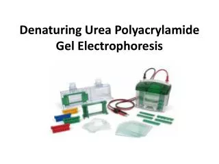
Experiment 7 . SDS- Polyacrylamide Gel Electrophoresis. BCH 333[practical] . Objectives : To be familiar with SDS-PAGE protocols (Linear slab gel ). SDS- Polyacrylamide Gel Electrophoresis.

E N D
Experiment 7 SDS-Polyacrylamide Gel Electrophoresis BCH 333[practical]
Objectives: To be familiar with SDS-PAGE protocols (Linear slab gel).
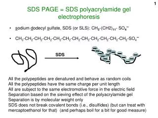
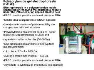
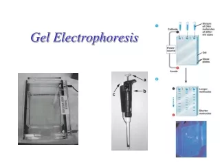
SDS-Polyacrylamide Gel Electrophoresis -Sodium DodecylSulfate-PolyacrylAmide gel Electrophoresis (SDS-PAGE) is a low-cost, reproducible and rapid method for qualifying, comparing and characterizing proteins[e.g. determining MW of proteins], checking purity of protein samples. -This method separates proteins based primarily on their molecular weights. -In general, fractionation by gel electrophoresis is based on differences in size, shape and net charge of macromolecules.[in molecules native condition using native gel electrophoresis].
-Systems where you separate proteins under native conditions cannot Distinguish between these effects and therefore proteins of different sizes may have the same mobility in native gels. -In SDS-PAGE this problem is overcome by the introduction of an anionic detergent SDS which binds strongly to most proteins. -When hot SDS is added to a protein all non-covalent bonds[?] are disrupted and the proteins acquire a negative net charge. -A Concurrent treatment with a disulfide reducing agent such as β-mercaptoethanol or DTT (dithiothreitol) further breaks down the macromolecules into their subunits.
The electrophoretic Mobility of the molecules is now considered to be a function of their sizes i.e. the migration of the SDS-treated proteins towards the anode[+] is aversely proportional to the logarithms of their molecular weights, or more simply expressed: Small proteins migrate faster through the gel. [Compare this with the situation in gel filtration.]
2-Polyacrylamide gel: • - The polyacrylamide gel is formed by co-polymerization of acrylamide and a cross-linking • By N,N’-methylene-bis-acrylamide. • To polymerize the gel a system consisting of ammonium persilfate (initiator) and tetramethylene ethylene diamin (TEMED) is added[catalyst].
Principle: • In denaturing protein electrophoresis, the addition of SDS to the electrophoresis buffer uniformly coats the proteins with negative charges, equalizing the charge to mass ratio for all proteins. • -A Concurrent treatment with a disulfide reducing agent such as β-mercaptoethanol or DTT (dithiothreitol) further breaks down the macromolecules into their subunits. • -So, the proteins samples are having uniformed structure and charge the separation will depend on their molecular weight only. • -Small proteins migrate faster through the gel under the influence of the applied electric field. • Note: The number of SDS molecules that bind is proportional to the size of the protein, thereby in the electrical field, protein molecules move towards the anode (+) and separated only according to their molecular weight.
graph of log MW vs. Rf is sigmoidal, it is nearly linear for a range of molecular weights excluding very small and very large M wt (Figure has been taken from http://www.nationaldiagnostics.com/article_info.php/articles_id/55)
-In practice the proportionality of log(MW) vs. Rmholds true for most proteins, provided they are fully denatured, and provided the gel percentage has been chosen to match the molecular weight range of the sample. -In fact, the actual plot of log(MW) vs. Rf is sigmoidal( previous Figure), because at high MW, the sieving effect of the matrix is so large that molecules are unable to penetrate the gel, while at low MW, the sieving effect is negligible, and proteins migrate almost at their free mobility, which in SDS is independent of MW.
SDS-Polyacrylamide Gel Electrophoresis Buffers and solutions: 1- SDS-PAGE, Running buffer (5x) pH 8.4:[?] Tris Glycine SDS Made up to 1L with distilled water. 2-SDS-PAGE, disruption buffer:[?] 10% (w/v) SDS [?] 1M Tris/HCl, pH 6.8 Glycerol [?] β-Mercaptoethanol [?] Bromophenol blue [?] with distilled water
3-SDS-PAGE, Stain: [?] Glacial acetic acid Methanol Coomassie brilliant blue R[?] Made up to 1L with distilled water. 4-SDS-PAGE, de-stain: Glacial acetic acid Methanol Made up to 1L with distilled water.
5-Sample preparation: 40µl of protein sample + 10 µl of disruption buffer boil the mixture 3minets at 99C̊ . 6-Separation gel contents:
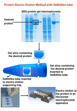
Applications: 1. To detect the purity of the protein 2. Determine of protein molecular weight http://www.youtube.com/watch?v=EDi_n_0NiF4