Gel Electrophoresis
230 likes | 552 Vues
Gel Electrophoresis. By: Tasnuva Jhileek Dr. Francine Norflus Biotechnology. What is Gel Electrophoresis?. A procedure that separates molecules based on size and charge. Uses an electric field Molecules move through gel made of agar or polyacrylamide. Simple Procedure.

Gel Electrophoresis
E N D
Presentation Transcript
Gel Electrophoresis By: TasnuvaJhileek Dr. Francine Norflus Biotechnology
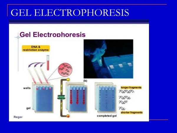
What is Gel Electrophoresis? • A procedure that separates molecules based on size and charge. • Uses an electric field • Molecules move through gel made of agar or polyacrylamide
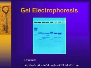
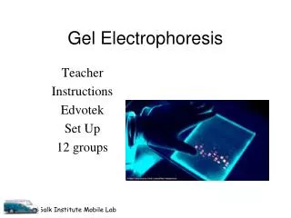
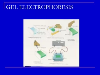
Simple Procedure • Molecules are dispensed into a well in the gel material. • Gel is placed in an electrophoresis apparatus • Electric current is applied to the contents of the apparatus • In the gel: • Larger molecules move more slowly • Smaller molecules move faster • The different sized molecules form distinct bands on the gel.
Types of Gel • Agarose – DNA and protein • Polyacrylamide – Protein and DNA • Starch - protein
Agarose Gel • Easy to cast, handle, store or dispose • Used to separate DNA fragments ranging from 50 base pair to several megabases (millions of bases). • Distance between DNA bands is determined by the percent agarose in the gel. • 0.7% (large 5–10kb DNA fragments) and 2% (small 0.2–1kb fragments) agarose. • Low percentage gels - very weak and may break • High percentage gels - brittle and don’t set evenly
Polyacrylamide • used for separating proteins ranging in size from 5 to 2,000 kDa - Uniform pore size • Pore size is controlled by controlling the concentrations of acrylamide and bis-acrylamide powder used in creating a gel. • used to separate different proteins or isoforms of the same protein into separate bands and small DNA • gels are made in 6%, 8%, 10%, 12% or 15%. • percentage chosen depends on the size of the protein • smaller weight = higher gel
Starch • Partially hydrolysed potato starch – protein electrophoresis • Non-denatured proteins are separated according to charge and size. • They are visualised using Napthal Black or Amido Black staining. • Typical starch gel concentrations are 5% to 10%
Nucleic Acid Electrophoresis • analytical technique used to separate DNA or RNA fragments by size. • an electric field induces the nucleic acids to migrate toward the anode • smaller fragments end up nearer to the anode • DNA is frequently cut into smaller fragments using a DNA restriction endonuclease (or restriction enzyme) or even PCR • Fragment size determination is typically done by comparison to commercially available DNA markers containing linear DNA fragments of known length • agarose (for relatively long DNA molecules) and polyacrylamide (for high resolution of short DNA molecules
Protein Electrophoresis • proteins, unlike nucleic acids, can have varying charges and complex shapes • proteins are usually denatured in the presence of a detergent such as sodium dodecyl sulfate/sodium dodecyl phosphate (SDS/SDP) • the rate at which the resulting SDS coated proteins migrate in the gel is relative only to its size and not its charge or shape.
Protein Electrophoresis cont. • Proteins are usually analyzed by sodium dodecyl sulfate polyacrylamide gel electrophoresis (SDS-PAGE), by native gel electrophoresis, by quantitative preparative native continuous polyacrylamide gel electrophoresis (QPNC-PAGE), or by 2-D electrophoresis.
Gel Condition • Denature - the native structure of macromolecules that are run within the gel is not maintained. • denatures the native structure of a protein • Native - separation method typically used in proteomics and metallomics. • does not use a charged denaturing agent.
Buffers • nucleic acids - Tris/Acetate/EDTA (TAE), Tris/Borate/EDTA (TBE). • TAE - lowest buffering capacity but best resolution for larger DNA. This means a lower voltage and more time, but a better product. • Xylene cyanol and Bromophenol blue are common dyes found in loading buffers
Visualization • DNA may be visualized using ethidium bromide • SYBR Green I – more expensive, quicker, safer
Ethidium Bromide • light sensitive and is stored in a brown bottle covered in tin foil (aluminum foil). • fluoresces under UV light when intercalated into the major groove of DNA (or RNA) • any band containing more than ~20 ng DNA becomes distinctly visible • protein may be visualised using silver stain or Coomassie Brilliant Blue dye. • Mutagen • ALWAYS wear gloves
Areas of Application • used in forensics, molecular biology, genetics, microbiology and biochemistry. • results can be analyzed quantitatively by visualizing the gel with UV light and a gel imaging device • the intensity of the band or spot of interest is measured and compared against standard or markers loaded on the same gel. • the measurement and analysis are mostly done with specialized software.
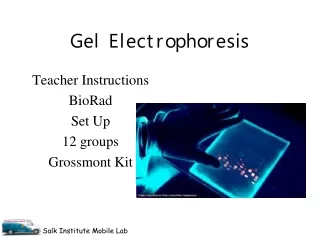
Lab Procedure • Make Ethidium Bromide • Make buffer • Add agarose to buffer and microwave • Pour gel after setting comb • Pour remaining buffer + EtBr in apparatus • Load gel (Black to Red 60 – 100 V) • Run gel • Kodak Moment • Discard waste product safely
Making EtBr • 1 g of Ethidium Bromide in 100 ml of dH20 in DARK colored bottle of transparent glass bottle wrapped in aluminum foil. • Dissolve completely (use Stir bar) • Use caution • Wear gloves at all times • Discard all EtBr related waste to biohazard
Making Buffer • Calculation: Total of 300 ml of solution • 15 ml of TBE + 285 ml dH2O
Making Gel • Calculations: 0.8% agarose gel in 100 ml of solution • Mix 0.8 g of agarose gel in 100 ml of buffer solution. • Microwave until agarose dissolves completely in buffer. • Let it cool (not solidify). • Pour!
Preparing the DNA sample • 10 microliter DNA + 2 microliter tracking dye in microcentrifuge tube. • Ladder: 1 microliter concentrated ladder per 9 microliter sterile water • Load all contents in separate wells in gel.
Setting up the apparatus • 5 microliterEthidium Bromide + 100 microliter TBE solution in electrophoresis apparatus • Plug apparatus into the electric outlet • Put gel in the apparatus (wells being in the black side) • DNA will travel from Black to Red • Set V to 60 or 100 • Run DNA for about 30-40 minutes
What is the safe way to discard the waste? • Put all EtBr related waste in the biohazard bin. • Discard buffer from the apparatus into biohazard bottle. • Wash gel in sink before throwing in the trash